Features of diffuse liver diseases: why hepatitis is so insidious
Viral hepatitis along with
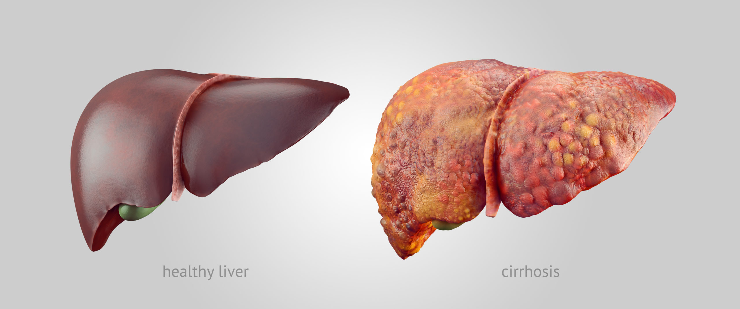 Healthy liver / Liver with cirrhosis
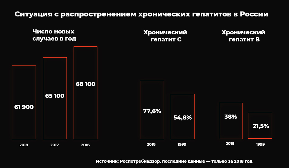
360 and 180 million people worldwide are infectedhepatitis B and C, respectively. 500–700 thousand people die annually from hepatitis B and more than 350 thousand patients die from complications of hepatitis C. High levels of incidence of chronic forms of viral hepatitis continue to be recorded in Russia - in 2018 they were registered in 61.9 thousand people . In addition, according to the departments of Rospotrebnadzor for the constituent entities of the Russian Federation, from 2009 to 2017 there were many cases of acute hepatitis C with unknown routes of transmission of the pathogen, which indicates an insufficiently effective epidemiological investigation of its foci.
Diffuse liver disease often developsimperceptibly and asymptomatic. Their origin can be of a different nature, so they affect people regardless of gender, age and the presence of concomitant diseases. Moreover, many of them cause parenchymal fibrosis, that is, the proliferation of connective tissue with the appearance of cicatricial changes. This condition can lead to cirrhosis - the replacement of liver tissue with connective tissue. Liver cirrhosis is deadly.
The most effective prevention tacticliver cirrhosis - identification of fibrosis in the initial stages. On the one hand, fibrosis is a consequence of the disease, not its cause. However, treatment of the early stages of fibrosis has shown good results. Even for those people who do not have risk factors for such diseases, it is recommended to be examined at least once a year. Keeping your liver healthy is very important. The function of a number of other organs and systems depends on its work: the nervous system, kidneys and lungs.
A little about the origins of hepatology
Scientists have begun to take an interest in conditionsliver back in the VI-V centuries BC. So, the ancient Greek philosopher Diogenes described the large vessels of the liver and introduced the concept of "hepatitis". And the ancient Greek healer, physician and philosopher Hippocrates noticed the connection of jaundice and ascites (dropsy) with liver diseases and was the first to comment on cirrhosis. In antiquity and for a very long time, palpation of the patient's abdomen remained the main method of diagnosing such diseases.
The founder of evidence-based hepatology -branch of medicine that studies diseases of the liver and biliary tract - became the ancient Roman physician Galen. He systematized a huge amount of medical knowledge accumulated since the beginning of the 5th century AD, and supplemented it with his own developments, and also created an entire medical system, which later became the basis for the development of this science. Galen first presented the detailed anatomical structure of the liver, correctly understood its main functions, identified four types of jaundice and proved that the liver is the central energy organ that supports the work of the heart.
In modern hepatology, the first breakthrough happenedin 1888, when the Russian scientist Sergei Petrovich Botkin told the world about a new infectious disease caused by the virus, which was later called the hepatitis virus, and also confirmed the relationship of this virus and liver cirrhosis.
To date, scientists have identified severalvarieties of the hepatitis virus, which differ from each other in many ways, including methods of transmission. However, they all have a significant impact on the liver and human health in general.
Diagnostics of the liver in the 20th century: the emergence of biopsy
The 20th century was marked by a number of significant events inhepatology. In 1963, the first donor liver transplant was performed. In the 1970s and 1980s, scientists discovered various hepatitis viruses and created their classification in 1994. And in the 1950s, thanks to the contribution to science of Sheila Sherlock, a hepatologist from the UK, biopsy became the main method for diagnosing liver diseases.
A biopsy is the removal of a small piece of tissueorgan (biopsy) to establish or clarify the diagnosis. The procedure is performed using special hollow needles. In a liver puncture biopsy, the doctor makes a puncture in the patient's abdomen or chest. The first case of the use of biopsy in medical practice was recorded in 1883 - the operation was carried out by the German doctor Paul Ehrlich. In addition to a puncture biopsy, since the 70s of the last century, doctors began to carry out the procedure transvenously, that is, to insert a needle into the liver through blood vessels.
A tissue sample obtained with a needle is sent to a laboratory where it is examined under a microscope. Based on the data obtained, the doctor can make a diagnosis.
Biopsy remains the most commona method for detecting liver diseases - it is called the “gold standard” of diagnosis. But it also has a significant drawback: it is invasive, that is, during the procedure the needle injures human tissue, including the skin and mucous membranes. The invasiveness of the procedure creates a risk of complications in the form of bleeding, injuries to the lungs and gallbladder, and peritonitis. In such cases, the consequence of a biopsy can even be fatal.
The 21st century with its achievements in instrumentaldiagnostics and imaging technologies has led me to wonder whether it is always appropriate to perform a biopsy to make a diagnosis. The procedure is not only painful for the patient and fraught with complications, but also may be ineffective as a method of assessing the stage of fibrosis. The fact is that there is a phenomenon of fabric “sample error”. If the biopsy is not representative enough, then the changes that a morphologist detects under a microscope may not fully reflect the state of the organ. In 2008, Professor Thierry Poinarou from Paris called biopsy the “imperfect gold standard.”
XXI century: shear wave elastography - a new standard for diagnosing liver diseases
Through the development of non-invasive techniquesdiagnostics in the 21st century, doctors manage to ensure the comfort and safety of patients during liver examinations. In European countries, shear wave elastography is now increasingly used.
Elastography is a modern technologydesigned to assess the degree of liver stiffness using ultrasonic waves. Today this technology is so ingrained in the world hepatological practice that it is reflected in the practical recommendations of the European (EASL), Asia-Pacific (APASL) and Latin American Association (ALEH) for the study of the liver. According to Giovanna Ferraioli, a professor at the Pavia University School of Medicine (Italy), a recognized world expert in ultrasound diagnostics of liver fibrosis, elastography is indispensable for examining patients with cirrhosis, people with severe or severe fibrosis who need immediate treatment, and when patients refuse from the biopsy procedure. The expert claims that elastography helps to reduce the time to assess the condition of the organ and make difficult decisions about the treatment regimen.
Patients in many countries have a positive attitudeto elastography, because the method is completely safe and painless. In addition, hospitalization is not required for the procedure, the results are given almost instantly. The research itself takes no more than 20 minutes. The only requirement is fasting for 4-6 hours before the procedure and obligatory rest for 10-20 minutes before it. The position of the patient is lying on his back with his right hand behind his head (this is necessary to expand the intercostal spaces, where the doctor places the ultrasound machine sensor).
The undoubted advantage of elastography for the doctor isthis is the completeness of information after just one procedure. The study provides not only data on the elasticity of the liver tissue, but also the gray-scale picture of the organ, as well as the parameters of blood flow in the portal vein system and the size of the vessels. All this helps specialists to correctly diagnose. In contrast to biopsy, shear wave elastography allows assessing the elasticity of not one limited area, but the entire field available for research. Given that fibrosis can spread and affect the liver in a mosaic manner, this fact is important to take into account.
Shear wave elastography in Russia
Shear wave elastography in ourthe country is not yet widespread enough. In accordance with the official recommendations of the Russian Society for the Study of the Liver (ROPIP) and the Russian Gastroenterological Association (RGA) from 2017, preference should still be given to biopsy, which is considered "a widely available and safe method for assessing morphological changes in the liver." Non-invasive methods for assessing fibrosis are cited as possible alternatives.
A major step towards the spread of elastography inin our country - the development of clinical guidelines that standardize the assessment of liver stiffness. Doctors need such guidelines as much as they need technological equipment. The document has already been prepared and published by the Russian Association of Ultrasound Diagnostics in Medicine (RASUDM) together with the Department of Ultrasound Diagnostics of the University of Pavia (Italy).
Values obtained during the procedureelastography are not least dependent on the equipment used. The elastography option is available on the EPIQ Series, Affiniti 70 series ultrasound units and on systems equipped with the eL18-4 high frequency array transducer. It is important to emphasize that the functionality of these devices is not exclusively limited to liver studies. The sensor can be used to assess various organs and body systems, including the mammary gland, superficial organs, musculoskeletal system, vessels and organs of the abdominal cavity. In addition, it is suitable for pediatric research and prenatal diagnosis.
Today you can study the experience of using the methodelastography in some medical institutions. For example, the procedure has been used for more than three years in Vladivostok, at the Asklepius multidisciplinary medical center. Denis Glushenko, head of the clinic’s ultrasound diagnostics department, says that the center uses elastography every day. 85% of studies are carried out on patients with hepatitis C, about 10% on people with hepatitis B and about 5% on hepatitis of unknown etiology. The expert said that elastography is easy to perform, but requires strict adherence to the technique in order to avoid diagnostic errors.
Since 2018, shear wave elastography of the liverintroduced into daily clinical practice in Barnaul. During this time, more than a thousand studies have been carried out. Lyudmila Shulgina, Doctor of Medical Sciences, Professor of the Altai Medical Institute of Postgraduate Education, Head of the Department of Functional Diagnostics of KGBUZ "Regional Clinical Hospital", shares two years of experience in using the method on an outpatient basis and in a gastroenterological hospital and notes that in order to obtain reliable results of elastography, careful observance of methodological principles and adequate staff training. In more than 90% of cases, people with chronic viral hepatitis B and C were sent to the institution. According to the doctor, a significant advantage of the method is its non-invasiveness, lack of complex preparation, speed of implementation, and the possibility of an unlimited number of repeated studies. The introduction of the elastography method made it possible to more reasonably choose the treatment tactics both in the initial manifestations of fibrosis and in difficult situations, in the presence of severe fibrosis and cirrhosis of the liver.
Shear wave elastography is called the future.liver diagnostics. It gives patients peace of mind and comfort, and doctors more confidence in making a diagnosis. Today, on World Hepatitis Day, it is high time for every person to remember how important it is to take care of their health and undergo a timely examination of vital organs. Moreover, modern technologies make it possible to do this quickly, safely and painlessly.
See also:
See what Hubble's successor telescope can see in space
The history of the origin of "strange" meteorites is revealed: they fell to Earth in the 60s
NASA explains why a meteorite from Mars will have to be returned back to the planet