An innovative method called HiP-CT, which uses X-rays produced by one of the
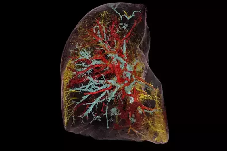
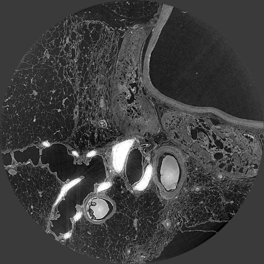
One of the first pictures taken withHiP-CT, is a snapshot of the lung of a person killed by COVID-19, which has already allowed doctors to figure out new details about how covid interferes with and disrupts the process of oxygenation of the blood.
The key point of HiP-CT technology isthe use of the ESRF synchrotron particle accelerator, which, after modernizations, has become the brightest source of X-ray radiation in the world today.
A group of researchers from the UniversityCollege London has already launched a project called the Human Organ Atlas, which aims to fill the existing gaps in our knowledge of the anatomy of our bodies. This project is being implemented as a publicly available online resource, which already provides high-quality images of several key organs of the human body, including the brain, kidneys, heart and spleen.
In the future, in the processing of data from the Human Organ projectAtlas will use artificial intelligence technologies that will take HiP-CT, MRI and CT scan data, and combine it all into a single set, providing physicians with a level of detail unattainable today.
Source: New Atlas