Researchers from the University of Bologna have developed a technique for analyzing archaeological bones, which
The scientists used hyperspectralnear-infrared imaging and a chemometric model to generate chemical images of the distribution of collagen in ancient bones. This model quantifies collagen in each pixel and thus provides a chemical mapping of collagen content.
Hyperspectral Near Infrared Camerarange used by the scientists is a line scan system. It collects reflected radiation data with wavelengths ranging from 1,000 to 2,500 nm. This analysis lasts only a few minutes and does not destroy the sample. On the finished map, scientists can find areas that contain enough collagen for deeper analysis.
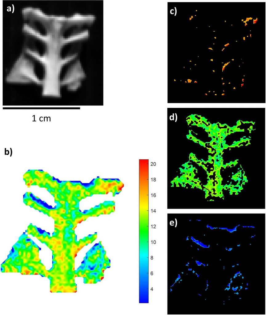 Scan results of one of the samples: the color map shows the percentage of collagen. Image: Cristina Malegori et al., Communications Chemistry
Archaeological bones can provide a lotinformation about the lives of ancient peoples: what they ate, their reproductive habits, their illnesses and the migrations they undertook. But the possibility of a full-fledged analysis depends on how much collagen is preserved in a particular sample.
due to diagenetic changes in collagenover time, a large initial weight of Paleolithic bones (more than 500 mg of bone material) is needed to extract a sufficient amount of material for analysis. Searching for the right zone in the blind means spending a lot of time, money and, most importantly, ruining valuable samples. Obtaining preliminary information about the distribution of collagen solves this problem.
Read more:
Look at the highest resolution map of Mars: 110,000 frames and 5.7 trillion pixels
"Sea" of quarks inside one proton: what does an elementary particle consist of
New image of Hubble puzzled scientists