Unlike existing brain imaging methods - MRI, CT or PET - the technology can be used to
Researchers are confident that their technology willsafe because sound waves are already used for ultrasound scanning, and the principle they offer uses similar sound frequencies. Ultrasound cannot easily penetrate the bone, while a new device designed to be worn in the form of a helmet is able to overcome this barrier.
The new approach is of particular value topatients examined for stroke, the second leading cause of death and the most common cause of neurological disability in adults. It is in the case of a stroke that fast, universally applicable and high-quality imaging is needed. Of particular interest is the fact that the same technology is used in monitoring seismic activity.
Dr. Louis Guash of the Imperial Department of Sciencesays about the Earth and engineering sciences: "The visualization technique, which has already revolutionized one area - seismic processing, can now radically change another - brain visualization."
Professor Brian Williams, Director of the CenterUniversity of California Biomedical Research adds: “This is an extraordinarily important advancement in brain imaging that has great potential for providing affordable research in routine clinical practice - for assessing injuries due to head injury, stroke, and other brain diseases.”
Scientists use seismic data andA computational algorithm called full waveform inversion to map the interior of the earth. Seismic data from earthquake detectors (seismometers) are included in algorithms that extract three-dimensional images of the earth's crust. They can be used to predict earthquakes and search for oil and gas reservoirs.
This approach has been adapted to medicalvisualization, having developed a method that uses sound waves with the ultimate goal of obtaining high-resolution images of the brain. The developers designed a helmet equipped with many acoustic transducers, each of which sends sound waves through the skull. The energy of ultrasound, which propagates through the head, is recorded and supplied through a helmet to a computer. Then the same algorithms are used to analyze the reverberation of sound throughout the skull, creating a three-dimensional image.
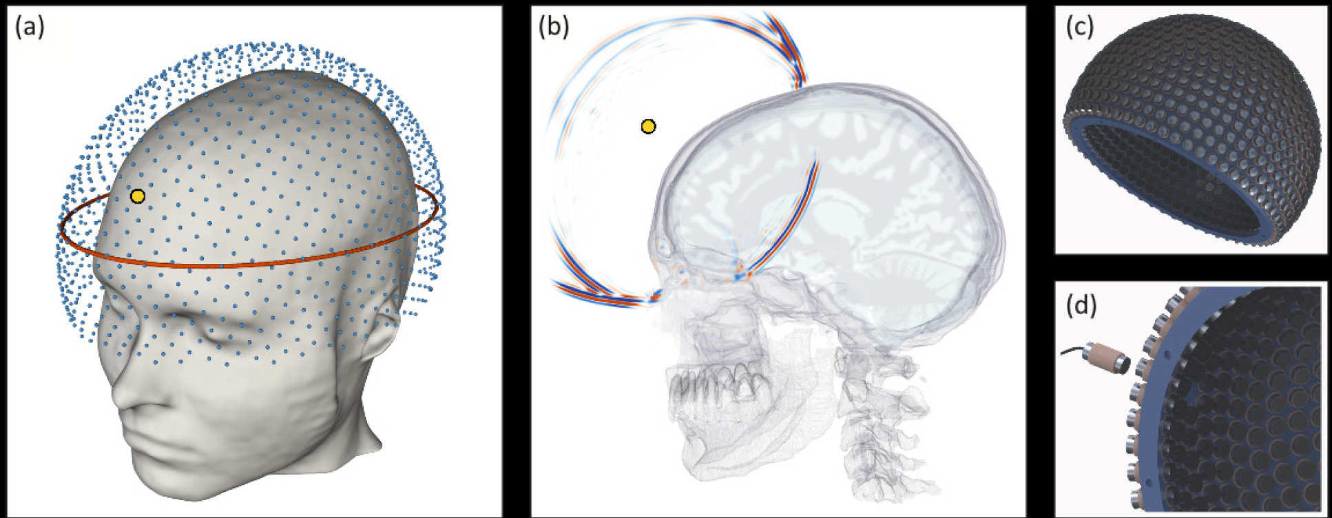
Researchers tested their helmet on healthyvolunteer and found that the quality of the recorded signals was sufficient for the algorithm for generating a detailed image, and they are sure that the scattered energy from the brain will be interpreted. Using computer simulations, they also found that they could receive high-resolution images with sufficiently low frequencies of sound to penetrate the skull with safe intensity.
They also created detailed computersimulations based on the properties of various types of tissue of the human brain to establish that sound waves will be effective in imaging the brain in high resolution.
Dr. Guash adds: “This is the first time that geophysical algorithms are used to visualize a human skull. Our joint multidisciplinary team of geophysicists, bioengineers and neurologists uses them to create a safe, cheap and portable method for generating three-dimensional ultrasound images of the human brain. ”