An international team of paleopathologists and medical experts examined the children's remains,
The researchers analyzed the mummifiedthe remains of 21 children aged 1 to 14. Mummies belonged to different eras and were found in different burials of Ancient Egypt. As a result of mummification, the remains of children were better preserved than those of those who were simply buried. Scientists have used CT scans to study children's skeletons without damaging museum exhibits.
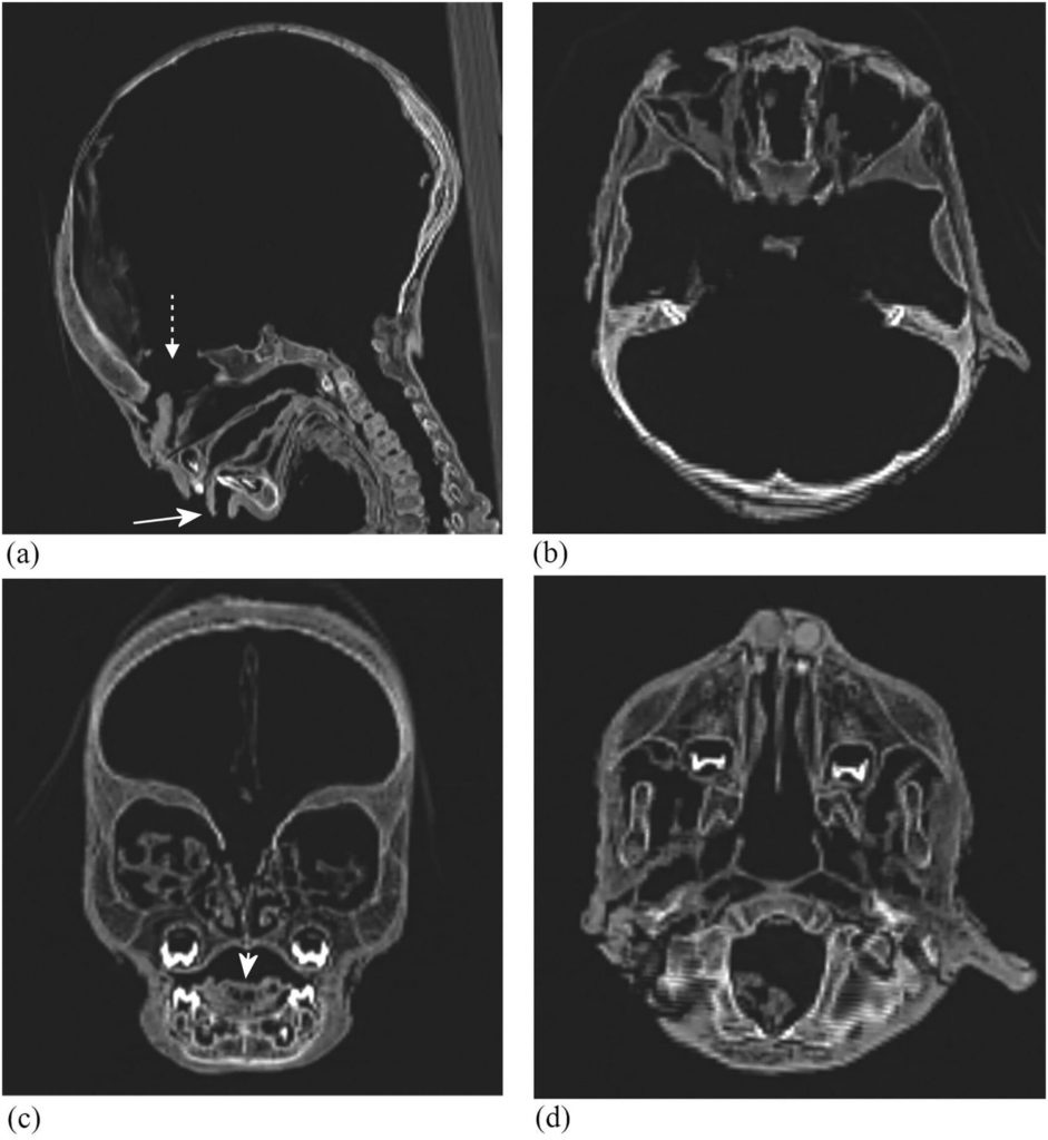
The analysis showed that a third of the children studied had a pathological increase in the bones of the cranial vault. This is a typical sign of chronic hemolytic anemia and iron deficiency anemia.
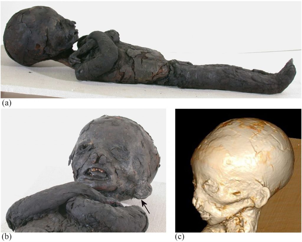 The mummified remains of one of the children andreconstructed skull shape (left), thickening of the frontal bones on CT scans (right). Images: Stephanie Panzer et al., International Journal of Osteoarchaeology
Anemia is a condition in which the amounterythrocytes or the concentration of hemoglobin in them falls below normal and as a result, the ability of the blood to carry oxygen to the tissues of the body decreases. The most common causes of this disease include nutritional deficiencies, especially iron, and some infectious diseases. Children with anemia often suffer from gastrointestinal bleeding, inflammation, and chronic infections due to a weakened immune system.
Examination using computed tomographymummies does not allow to unequivocally identify the cause of the disease, as well as to determine whether it caused early death. But researchers believe that the most likely cause is a lack of nutrients, and the disease itself was at least one of the reasons that affected life expectancy.
Read more:
No longer a toy. What will the evolution of GPT and Midjourney lead to?
In space, no one will hear your cry: 7 action-packed books about the secrets of the universe
"Sea" of quarks inside one proton: what does an elementary particle consist of