Start regeneration
The World's First Successful Organ Transplanthas occurredIn 1954 he became a surgeon
Every year, an average of 100,800 are made worldwideorgan transplants. The most commonly transplanted transplants are kidneys (69,400 operations), liver (20,200), heart (5,400), lungs (2,400) and pancreas (2,400). Donation can be related and posthumous, when the desired organ is taken from a corpse. At the same time, in Russia, for example, the average waiting period for a donor kidney is 1.5-2 years. At the Sklifosovsky Research Institute, about 200 operations are performed annually, while there are approximately 500 people on the waiting list.
Nature has created cells in such a way that they always know what to do.They have the ability to regenerate, eachthe cell has such a potential. Skin cells are updated every two weeks, intestinal cells - for two weeks, and brain cells - every ten years. The problem is that we do not regenerate tissue when a disease, scar or damage occurs. At this point, regeneration stops, and regenerative medicine can help here. We take very small muscle tissue from the patient, then we process these cells and place them in the area where the damaged muscle is located. It can also be used for patients with burns: in this case we take a small sample of the patient’s skin, then we process the cells and simply apply them to the damaged areas with a spray. Moreover, if the patient is injured, you must first treat him, get rid of the infection and wait for the time to be ready for treatment.
Grow a new organ
Instead of cells, you can use the so-called substrate - a kind of building frame.Its materials are very similar to suture materials.They dissolve in a few months and are safe for humans and cells. We take a small sample of tissue from the patient, then we process those cells outside the body, culture them, use a scaffold to make them tubular, and implant it into the patient. The entire process takes approximately 30 days. The same applies to blood vessels. We put these cells on the material, then we train this organ. When the compression becomes what we need, the vessels are implanted in people. The most complex organ is a solid organ with blood circulation, like the heart, kidney and liver, because there are different types of tissues, and they all have so many blood vessels.
 Healthy Moscow
The easiest way to grow simple fabrics. In clinical practice, the method of skin regeneration using special hydrogels or cells of the patient is already used.
Gordana Wunyak-Novakovich at Columbia University grew a fragment of the skull bone, sowing the frame with stem cells.
At Johns Hopkins University, doctors removed frompatients ear and part of the skull affected by the tumor. Taking the cartilaginous tissue from the chest, blood vessels and skin, they grew a new ear on her hand, and then transplanted the artificial organ into place.
Successful experiments on the cultivation and transplantation of blood vessels were held at the universities of Gothenburg (Sweden) and Rice (USA). There are also examples of growing muscle, blood cells, bone marrow, and teeth.
Regarding the cultivation of complex organs,experiments are still being conducted mainly on animals. However, there are examples of successful transplants of artificially grown organs to people. For several years, Anthony Atala has been performing implantation operations on the bladder grown from the patient’s cells. Spanish surgeons transplanted a trachea grown on the basis of donor caracas.
Advanced Cell Technology in 2002grew a 5 cm long miniature kidney of a cow using cloning technology, taking cells from the animal’s ear. A kidney was implanted next to the main organs, and she began to successfully produce urine.
There is also positive experience in growing and transplanting livers into laboratory rats (University of Massachusetts) and lungs into pigs (University of Texas).
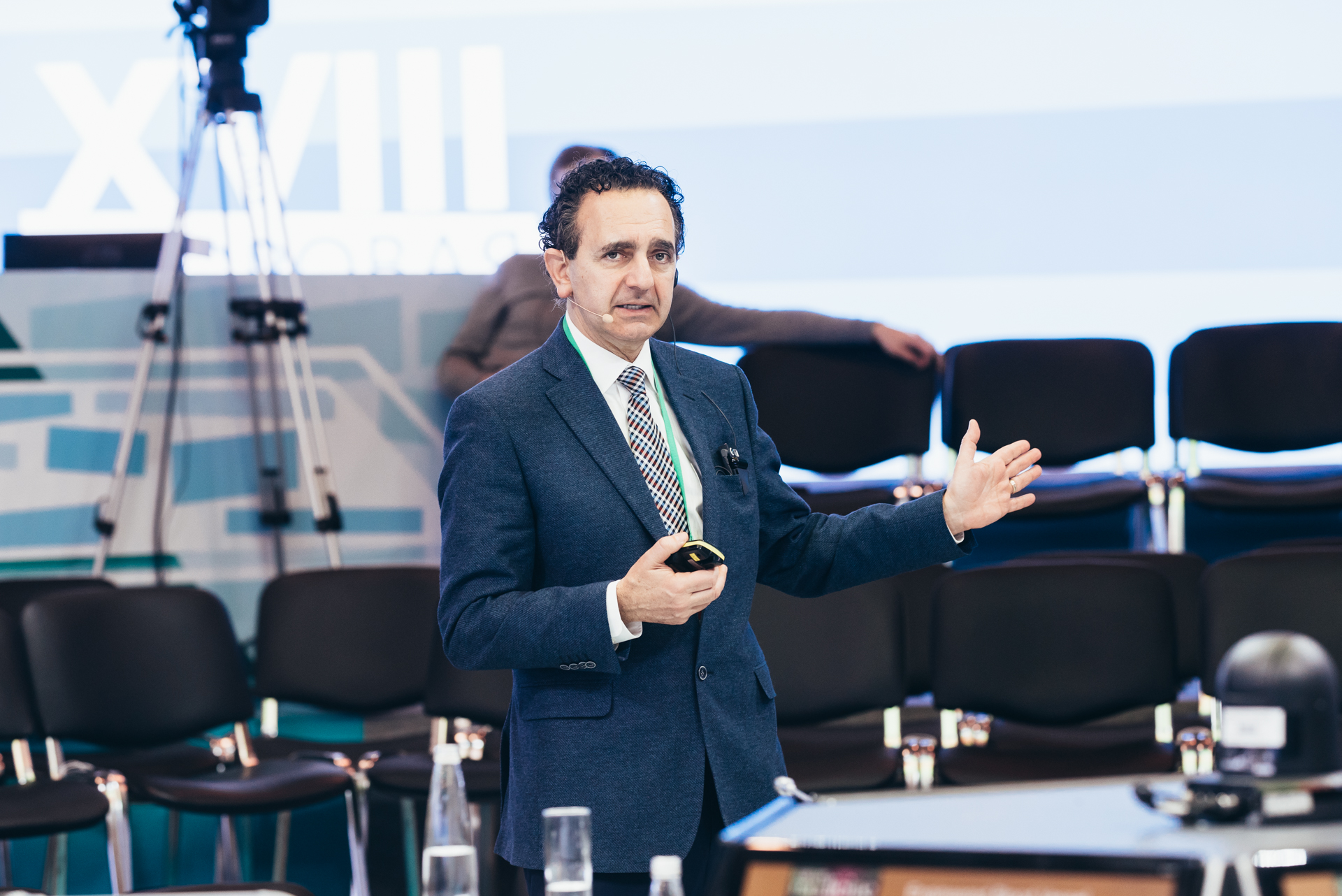
The vessels of the transplanted organs are very small.We already started this work 30 years ago, but wethere was no technology. They began to think about taking the organs of people who had died and using them again. They removed the liver from a deceased patient and, as it were, washed it from the inside. After two weeks, the liver still looked like a liver, but there were no cells inside. However, we were able to preserve the vascular tree, such as the skeleton of the liver. Then they took the patient's cells, grew them and placed them on this skeleton. We create tissue from the patient's body and thus treat him. Therefore there is no immune response at all. This is a very big advantage of regenerative medicine.
 Healthy Moscow
Even from a very bad organ, we can get good cells using a biopsy.But we cannot do this with geneticdiseases, because the defect will be in the entire tissue. Here are other technologies - we take cells from this patient, correct defects, how to heal these cells, and then work on the same strategy. So far, unfortunately, this is experimental, but there is still hope that genetic diseases can be treated.
We always trace the lives of our patients for at least 5–8 years after transplantation.We must make sure that everything will be fine, only then can we say that this technology worked and the transplanted organs are functioning normally.
Organ printing and drug testing
You can print a miniature heart, and within two hours it will already be beating.Six years ago, we started using 3D printing,because it was necessary to scale these technologies - before that we did everything manually. But organs obtained by printing did not have such integrity to be implanted into the body. Then we began to develop more specific printers that could create human tissue. And they worked on it for 14 years.
The first bioprinting experiments were conducted onOrdinary household 3D printers that have been upgraded in the field. In 2000, Thomas Boland set up Lexmark and HP machines so that DNA fragments could be printed on them, and in 2003 he patented the technology.
Now severalcompanies. Organovo's bioengineers have developed technology to print liver tissue. They also printed kidneys that remained functional for two weeks. So far, such bodies are used only for testing medical preparations, but the creators do not exclude that they will soon begin to develop equipment for printing donor organs.
Russian bioengineers from 3D Bioprinting Solutions developed the FABION 3D printer and conducted a successful experiment in printing the thyroid gland and transplanting its experimental mouse.
Designed by Sheffield University, Fripp Designs printers print eye prostheses. The same team is developing 3D printing technology for noses, ears and chins.
Some of the equipment is produced for our ownneeds of customers and is not intended for sale (FABION, Organovo’s NovoGen MMX). Prices for commercial bioprinters start from $10 thousand (BioBots) and €5 thousand (CELLINK Inkredible) to $200 thousand and above (EnvisionTEC’s 3D Bioplotter, RegenHU’s 3DDiscovery).
There are five interesting criteria for a 3D printer for printing organs.Firstly, they have very small nozzles, theycan reach up to 2 microns - this is 2% of the diameter of a human hair. Secondly, this printer gives us precision, we can lay out the cells where they are actually needed. The third is bioink, a liquid that passes through the nozzle. And then, when it becomes gelatin, it already functions as normal tissue. The next criterion is microchannels, they provide nutrition to the central part of the cells. Essentially, these are blood substitutes. And finally, software that allows you to have a three-dimensional image. In this way, we understand what is happening in the body and create the structure required for a given organ. To do this, we take digital data from the X-ray and use it to create a structure specifically for this defect in a particular patient.
 Healthy Moscow
We have two certified systems for printing human organs.They are FDA approved.Food and Drug Administration in the United States - Hi-Tech). Over the past six years, we have used a printer to create the so-called body-on-a-chip program. Now this is at the development stage, because it is necessary to ensure the viability of these organs, but in general we can make miniature lungs, heart, blood vessels and connect this whole system on chips. We can also create miniature organs the size of a pinhead and see how these organs respond to medications. For example, if a medicine speeds up your heart rate, it will speed up your heart rate in our miniature heart. Thus, it is possible to identify side effects of drugs that are not detected by other tests.
Avoid side effects
Using printed miniature organs, you can test the medicine.For example, the drug "Hismanal", 11 yearson the market all over the world. This antipsychotic has also been used as an antihistamine. After some time of its use, it became clear that many patients experience side effects associated with cardiac dysfunction. When the drug was tested on a cell, there were no problems; when it was tested on animals, there were no problems. When the first, second and third stage clinical studies were conducted, nothing happened. We took this medicine, used it on our organs on chips, and within a week it became clear that this medicine was toxic to the heart.
This is due to the fact that everyone reacts differently.for drugs, everything is genetically different, the same medicine will be processed differently. Everyone has a different diet, different living conditions, different health difficulties. This acts as a hindrance to understanding what the medicine actually does with the organs. And if we remove all these obstacles and see directly how the medicine acts on the organs, we can immediately detect toxicity.
Today we are developing a system that we call the “body on a chip”.It will particularly help reduce toxicity.medications - for example, to work with cancer patients. We can take a small cancer cell and grow it, and then test chemotherapy on a chip before giving that therapy to the patient. We had a patient with melanoma, he was on chemotherapy for six months, a lot of money was spent, and the tumor only grew. We tested a drug that had not been considered and the patient began receiving it. Two weeks later, the patient said for the first time that his condition was improving, and the doctor noted that the tumor was decreasing. Thus, it is very useful to test the medicine before giving it to the patient.
Stem cells: create what is not
When you need to grow an organ that the patient has never had or has lost, stem cells can be used.Usually, in order to grow a kidney, we takekidney cell, to grow the urethra, we take the cells of the urethra. But in the case of stem cells, we can take one that can become a cell in a lung, kidney, or blood vessel. There are two main types of stem cells. One of them is human embryonic. They are very powerful, grow and can turn into anything, but they can also form tumors, so it is very difficult to use them. On the other hand, if we talk about these cells in an adult, they can be fat or bone marrow cells, they will not form tumors, but they do not grow so well.
For the first time, the term "stem cell" usedGerman scientist Valentin Haacker at the end of the 19th century. In 1909, the Russian scientist Alexander Maximov suggested that there are cells in the body that remain unchanged, but at the right time they can change the program and turn into cells of a different type.
This theory was confirmed in the 60s.last century. Americans James Till and Ernest McCullough irradiated the mice with a lethal dose of radiation, and then transplanted them with a stem cell from a healthy individual. It turned out that in this way it is possible to restore blood and save mice from death. Since 1964, this method has been used in the treatment of blood cancer: patients first destroy their own blood cells, and then transplant healthy stem cells from the donor. The effectiveness of this method reaches 70–80%.
In 1981, Martin Evans and Matthew Kaufmanin parallel with Gail Martin, embryonic stem cells from mouse embryos were isolated. These cells could exist indefinitely outside the body without changing their properties, and when they get into certain conditions, for example, back into the body, turn into tissues.
In 1999, Science magazine ranked the discovery of stem cells as one of the three greatest discoveries in biology, after decoding the DNA and the Human Genome program.
For a long time it was believed that if a stem cellturned into a tissue cell, it is impossible to make it stem again. However, in 2006, the Japanese Xinya Yamanaka discovered a way to turn somatic cells back into stem cells. For this, in 2012, he received the Nobel Prize.
 Healthy Moscow
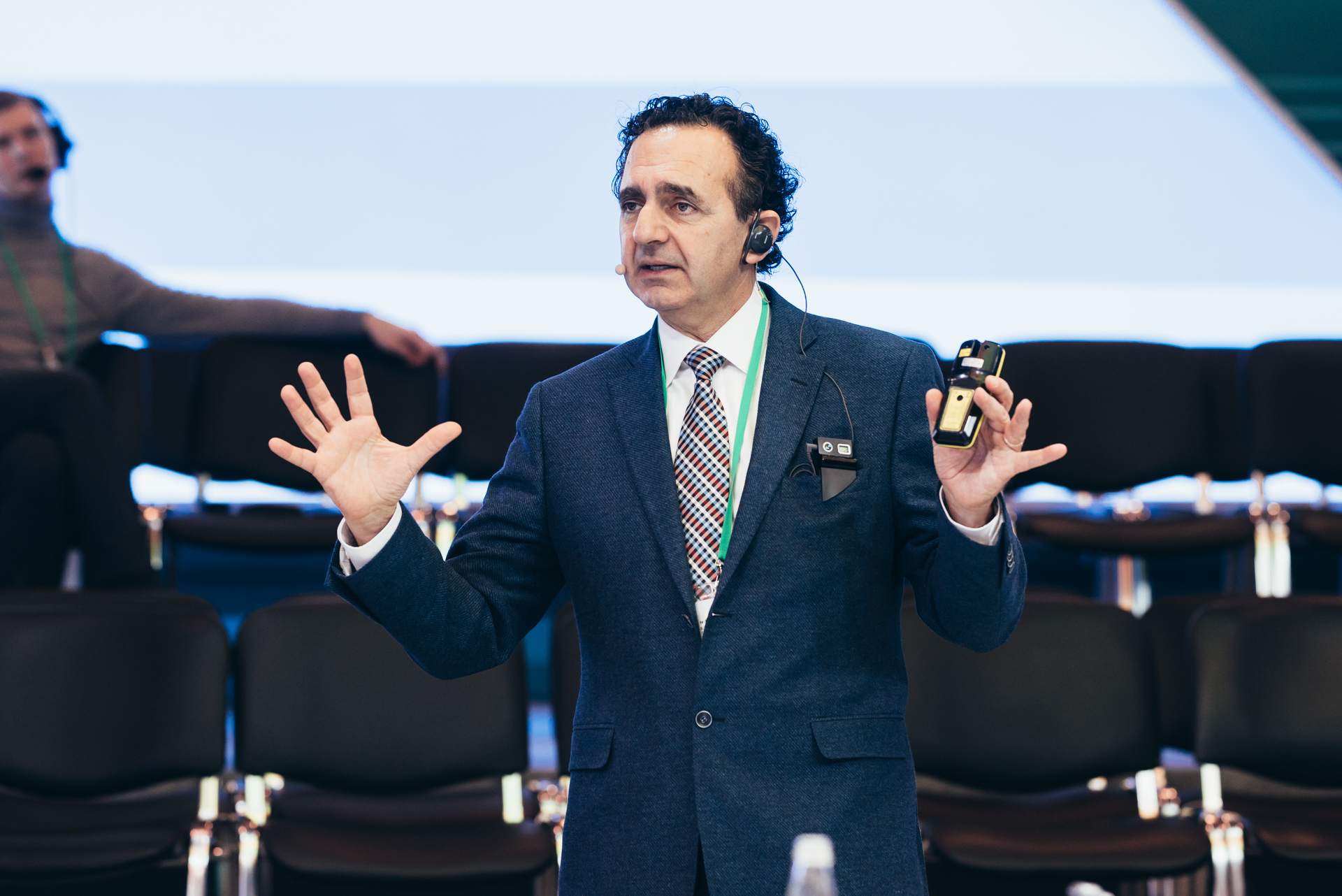
About 17 years ago, we started looking for an alternative source of stem cells.. Suggested that there is another type of stemcells present in the amniotic fluid and in the placenta with which the baby is surrounded in the uterus. And we found these very powerful stem cells. They will not form tumors and can turn into three main categories of tissue that form our body. These cells can be quickly grown to sufficient amounts. In this way, we avoid all the limitations of bone marrow cells and other types of cells. Now they are the subject of a number of clinical studies and so far are not used very widely.
I don't want you to think that all problems have already been solved and you can just print out organs.It will take decades for thesetechnology could evolve. This is very difficult, and it takes a lot of time for us to be able to develop the very recipe that will allow the technologies to work optimally. In addition, these are expensive technologies, it will be difficult to replicate them, but we can say for sure that they have potential. And for us, this is the promise of regenerative medicine - to make patients' lives better.