What happens to the brain in 30, 40 and 70 years
Our work concerns types of brain activity, but the focus is mine
The focus of our research was agethe differences are activity in the functioning of the brain in young and elderly subjects. This topic is important because of neurodegenerative diseases associated with aging (Alzheimer's disease and Parkinson's disease, dementia). There is no cure as such, but it is very important to identify the disease early and begin rehabilitation.
The brain undergoes a lot as we age.changes, like the whole body. They can be both structural and functional. Structural are changes in the functioning of an organ. The plasticity of the brain also changes—it is because of this that we learn, not only gain knowledge, but also motor skills. This property also suffers over time and affects both motor and cognitive functions.
How the brain in the elderly compensates for organ aging
The brain is an organ made up of severalshares. The motor cortex contains the most valuable information about how it processes movements. Therefore, according to the signals of the electrical activity of the brain, taken precisely from the motor cortex, it is possible to obtain a lot of results, to identify patterns associated with movement. Each of these areas is responsible for something different, and movements are not only processed in the motor cortex, they involve other areas depending on age.
 “The brain is an organ that consists of several lobes”
Electricity is taken from neurons - so they are each othercommunicate with a friend. It is transmitted through connections to other neurons and forms a pattern. There are also studies of chemical compounds that occur between neurons, they can also be studied.
Structural changes in the brain relate to volumewhite and gray matter. Gray is the substance of the brain, which consists of a body of neurons. This is the outer shell of the brain, the white matter is mostly inside and is covered with gray matter. There are no neurons in white matter, but there are connections between neurons - synapses.
On functional magnetic resonance imagingIn the brain of a healthy elderly patient and an Alzheimer's patient, you can see the degree of gray matter loss with age - this is normal. But in Alzheimer's disease, there are many red areas. This refers to the loss of gray matter by more than 4%, which is much more than with normal aging. The gray matter contains neurons, so the brain cannot function as it used to. And in elderly patients, losses do not exceed 2% - this is within the normal range.
White matter contains synapsesthere are no neurons in it, but it also decreases in volume with age. On the scans of a 25-year-old and a 75-year-old man, white areas indicate loss in white matter - more often they are lost due to aging. This is a normal process in which the brain rewires itself.
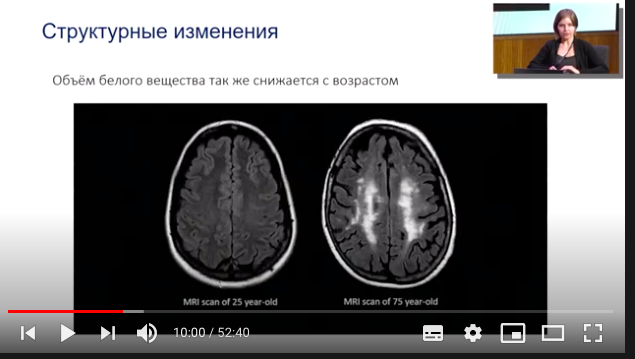 Scans of a man aged 25 and 75
When talking about functional changes, they havein view of changes in the network structures that are formed in the brain. The network structure can be built and detected by analyzing the electrical activity of the brain, which is what we are doing. The main marker of aging is the decay or decrease in the number of connections, but at the same time, there is an increase in connections between different networks. So the brain wants to compensate for the loss of connection in order to maintain its functionality. This is called the compensatory function.
On the network of functional connections between young and oldin a person without severe neurological diseases, the difference is immediately visible. In young people, the network of functional connections consists of four large clusters. At the same time, networks break up with age, the connections within each network become weaker, which leads to the network breaking up into several subnets. But, nevertheless, the connections between networks are much stronger than those of young people. This is precisely the illustration of the compensatory mechanism.
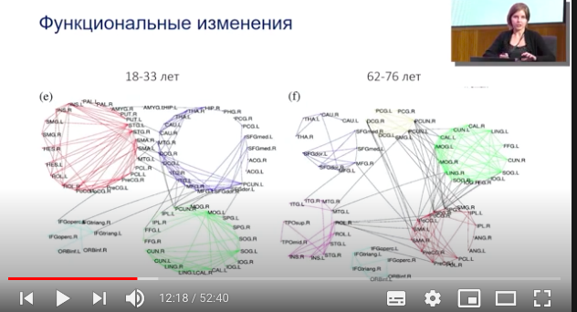 Functional changes in the brains of two patients
How the brain is examined
We have an experimental laboratory based onwhich we conduct experiments on recording electroencephalographic signals in various groups of subjects. They can be written for different purposes, but I will talk about motor functions.
The cap facilitates the insertion of the electrodes, whichread signals from different areas of the brain. Red squares and circles highlight those channels that enter the sensorimotor region. They carry the most significant information about how the brain is activated at the time of movement. At the same time, motor activity is a very loose concept, it is not only the execution of movements, but also their planning.
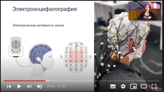 How is the electrical activity of the brain measured?
The patterns of these types of activity are quite similar, but they need to be able to distinguish. Therefore, we are engaged in the creation of a good classifier and brain-computer interface.
In experiments, we write a background entry, this is howcalled resting state recordings, during which a person sits with his hands folded calmly. His eyes are open, he is listening to some music, and we ask you not to think about anything in particular, so that there are no noises on the electroencephalogram records.
 Resting state entry
On the electroencephalogram recording, the numbers mean, inwhat part of the brain they are in. C3 is the left hemisphere, C4 is the right hemisphere. There is a well-known effect on the contralaterality of motor activity - if you move your left hand, the right hemisphere is activated, and vice versa. Therefore, it is important to write a complete arrangement.
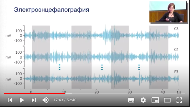 Electroencephalogram recording
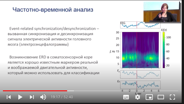
In the timeline from 0 to 40 seconds, three signals are highlightedfragments, where a person made movements - squeezing and unclenching his hand. Nothing is visible on raw signals. Therefore, it is necessary to apply signal analysis methods, and one of the most classical methods is time-frequency analysis.
 Time-Frequency Analysis
A bright picture is a surface, it containsinformation about the energies of each frequency range. Above is a fragment of the EEG signal, the red vertical line marks the moment when the sound signal passed - after it the person made a movement. The green stripe on the surface corresponds to the alpha rhythm. We measured the surface and calculated that the green bar is approximately in the range from 8 to 14 hertz, which corresponds to alpha rhythms.
Alpha rhythm is the basic rhythm of the brainwhich is responsible for a large number of tasks. In the context of motor activity, it also carries a very large amount of information about how the brain copes with our movements. For example, it is known that when a person makes a movement, the alpha rhythm loses energy, and this is called desynchronization of the alpha rhythm.
Age differences
The picture, where two curves are presented, is the average energies of the alpha rhythm. The green line is young subjects from 18 to 35 years old, the red stripe is elderly subjects aged 55–70 approximately.
On the left hand, the difference is not particularly obvious, butif you look at the right hand, you can see that the alpha rhythm after the signal, which is at zero, falls into both groups, the energy decreases, desynchronization occurs.
 Time-Frequency Analysis
But in young subjects, this desynchronizationlasts until the end of the movement and the alpha rhythm is restored after the person opens his hand. In older subjects, the alpha rhythm begins to return to zero soon after clenching his hand.
It is possible to measure, evaluate age differences andanother way. For example, the speed of a motor reaction. It is often said that old people have a reduced speed of reaction, movement, reaction, and coordination. But how does it happen?
Reaction time is how fast the neurons in your head are.begin to activate after a person has made a movement. We measured this for young people and for older subjects. Red squares indicate movement, reaction time for older subjects. Blue color - for young subjects. It turned out that in older people the reaction time is always longer than in young people for both the left and right hands. And in young subjects, the reaction time on the right hand is much lower than the reaction time on the left hand. And this is logical, because in our sample there are only right-handers.
We also observe the plasticity of the skill:the more a motor skill is trained, the faster it degrades with age. The skill of the left hand of right-handers is not particularly trained, so the degradation is slow, and the skill of the right hand is catching up with him. This leads to age ambidexterity, equal development of the functions of both hands. True, in the case of older people, unfortunately, this means that they are equally bad at making movements with both right and left.
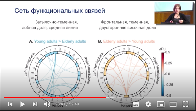
In the photo below, the blue picture is the links betweendifferent channels, they are stronger in young people than in older people. In the red picture, the connections that are stronger in older people than in young people - and you can see that they are different. In younger people, the connections are stronger in the occipito-parietal, frontal, and midline.
 Network of functional connections of two organs
In the elderly, the main activity shifts forward,in the frontal parietal zone, bilateral temporal lobe. This conclusion can be drawn from the analysis of the electroencephalogram recorded at the moment before the start of movement after the signal, in the interval when the signal sounded, and the person had not yet begun to move his hand. This segment contains a lot of important information about movement planning and response to a signal.
Defeat Alzheimer's
We can conclude that it decreases with agethe efficiency of these neural mechanisms that process motor activity. Also in the control group, based on which zones are activated in the elderly and in the young, motor memory is actively working in the latter.
Motor memory is how the brain remembersdifferent movements. And instead of performing the movement anew each time, he draws a resource from the motor memory, and this allows him to react faster. Athletes have a very developed motor memory because they train their reaction speed and motor skills.
Older people don’t have this, motor memoryworks worse, so each movement must be done again. Due to this, the speed of the motor reaction and the severity of desynchronization, which acts as a marker of motor activity, decreases.
 Two types of aging
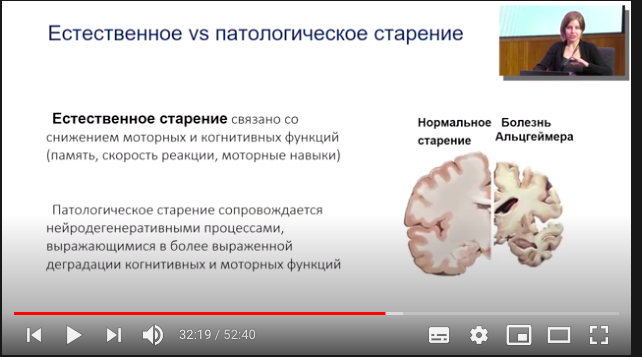
There is an opinion that for the elderly, diseasesAlzheimer's and dementia are normal. For some reason, it is believed that with age they develop that there is nothing pathological in this. But this is not so - they are called neurodegenerative for a reason.
With normal aging, the volume of white decreases andgray matter. But it decreases in a manageable way and not as dramatically as in Alzheimer's disease. This disease has devastating consequences for the brain: there is practically no gray matter, and the volume of white matter decreases. Unfortunately, there are currently no strategies for treating neurodegenerative diseases. An urgent task now is the development of methods for the early detection of signs of these diseases.
Read more:
Starlink signal hacked to be used as an alternative to GPS
NASA revealed the origin of Haumea - the most mysterious planet in the solar system
Physicists Break Standard Quantum Limit With 'Quantum Horror'