Researchers from the Catholic University of Korea and Seoul National University have developed
Light is repeatedly scattered in complexstructures such as living tissue. Because of this, the photons randomly change their direction several times as they pass through the tissue. As a result, during classical scanning, most of the electromagnetic waves are distorted, and the image is blurred.
For their work, scientists quantitativelyanalyzed how light and matter interact. In particular, the researchers developed a method to preferentially select singly scattered waves, using the fact that they have similar reflection shapes even when light hits from different angles.
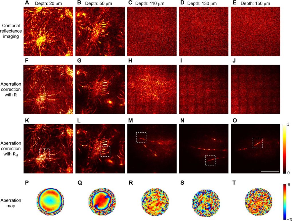 Images of the mouse cerebral cortex obtainedusing confocal reflective imaging (top row) and noise-corrected images (second and third rows). Image: Yonghyeon Jo et al., Science Advances
Using a complex algorithm and numerical operation,which analyzes distortion and interference between different wavelengths of light, researchers have developed a microscope that focuses more than 80 times more light energy onto nerve fibers than before. In this case, the device uses a probing beam in the visible wavelength range and is able to selectively process and suppress side signals.
In a series of experiments, scientists made severalimages of the brain of a mouse. The test results showed that, despite a large amount of noise and scattering, the device produces high-contrast images with a resolution of 412 nm.
Read more:
Look at a phenomenon that is simply impossible on Mars
A huge satellite can outshine all the stars and planets in the night sky
China says they will have fusion power in 6 years