The ability to remember and store memories for a few days or a lifetime is an important function
Understanding how the brain stores information andregulates which memories will remain for a long time, and which ones will disappear, will help develop methods for strengthening memory in those at risk of developing age-related disorders, and restore normal brain function after an injury.
How does memory work?
Various types of memory are created and storeddifferently and in different areas of the brain. Neuroscientists do not yet fully understand the intricacies of all processes, they continue to refine the details and discover new brain functions. However, it is known that autobiographical memories—memories of events experienced personally—begin to take shape in a part of the brain called the hippocampus in the hours and days following the event.
Neurons are the cells of the nervous system that communicate with each other.another through synapses. These are areas where two cells connect and exchange "information" through a tiny gap using chemical messages (neurotransmitters). Each neuron can be connected via synapses to thousands of others.
Interaction of neurons under a microscope. Video: UC Berkeley
One of the key properties of neurons is synapticplastic. This is the name given to the ability of synapses to strengthen or weaken over time in response to an increase or decrease in interaction activity. It is believed that long-term changes in the efficiency of synapses, depending on the frequency of "use", are important for learning, memory formation and neuronal development.
Neurons are constantly producing new proteins forremodeling of parts of the synapse, such as receptors for certain neurotransmitters. This allows nerve cells to selectively strengthen their connections with each other. As a result, a network is formed that encodes the memory. The more often a memory is “activated”, the stronger its neural network becomes. Such structures go beyond the hippocampus and form long-term memory in various parts of the brain.
Can you see memories?
At the end of the 19th century, scientists created the firstmicroscopes are powerful enough to identify individual neurons. By the middle of the next century, electron microscopes showed synaptic structures only a few tens of nanometers wide, and later, using two-photon microscopes, researchers observed how synaptic connections are formed in real time during the learning process.
One of the models that neuroscientists usefor working with memory it is an engram. This is the name given to the physical trace (neural network) of a particular memory in the brain. Engram cells are populations of neurons whose reactivation leads to individual memory retrieval.
Numerous researches in the field of geneticsmade it possible to visualize such engrams. For example, scientists have used viruses to inject a green fluorescent protein found in jellyfish into the brains of mice, causing neurons to glow as they learn. And by introducing the light-sensitive protein of algae, canalrhodopsin (ChR2), it is possible to artificially activate certain neurons, “turn off” or “start” certain engrams.
For example, researchers from MIT identifiedan engram that was formed in the brain of mice during the process of learning fear. Repeated artificial activation of this network of neurons using blue light caused the animals to “freeze,” a characteristic reaction to danger.
Another method of visualizing memories isfunctional magnetic resonance imaging (fMRI). This technology is based on the connection of neuronal activity with changes in blood flow in the brain. By observing how hemodynamics (blood movement) changes, researchers determine which areas of the brain are active at one time or another.
With this technology, for example,Researchers from the University of Oregon trained AI to recognize and reconstruct facial images that pop up in human memory. During the training process, participants were shown photographs of different people's faces, and a computer processed the fMRI data and generated patterns of brain activity characteristic of each photograph.
After that, when the participants were shown a newunknown AI photo, based on brain activity, the computer tried to reconstruct the face in the picture. Although it was far from being completely similar to the finished image, the artificial neural network accurately identified and recreated some features, and also reflected the subjective perception of certain features by a person, for example, skin color.
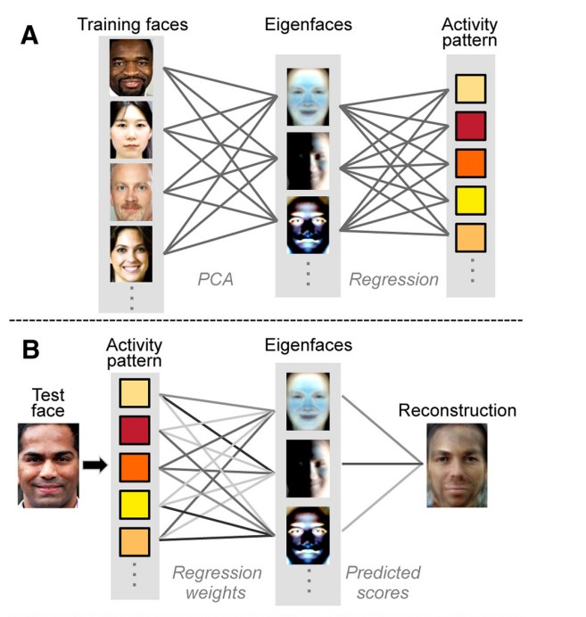 Experiment scheme: training (top) and reconstruction of an unknown image (bottom). Illustration: Hongmi Lee, Brice A. Kuhl, Journal of Neuroscience
Can memories be manipulated?
One of the ways to form "falsememories” in mice was demonstrated almost a decade ago by researchers at the Massachusetts Institute of Technology. The approach proposed by scientists is based on identifying engrams associated with certain events and activating them using optogenetics (controlling neurons with the help of light).
 Scheme of the experiment to create falsememories. The scientists read the pattern corresponding to environment A. They moved the animals to environment B, turned on the current, and in parallel with the help of light activated the neurons of the engram corresponding to environment A. When they were again placed in context A, they showed a false memory of fear for A (freezing is indicated by wavy lines ), where they were never electrocuted. At the same time, there were no changes in behavior in neutral environment C. Image: Steve Ramirez et al., Frontiers in Behavioral Neuroscience
Scientists have genetically modified mice toin order to introduce the gene encoding the protein canalrhodopsin (ChR2) into neurons. It is a light-sensitive protein that serves as a photoreceptor in unicellular green algae. The gene has been modified to trigger the expression of a fluorescent protein when the neuron is activated. This modification allowed the scientists to keep track of which neurons are active (fluoresce) during the learning process, as well as reactivate them using light.
During the experiment, scientists placedlaboratory mice into the first “room” and read the engram (neural network) that corresponded to the memories of this environment. After this, the animals were moved to the second environment, the neurons associated with the first “room” were activated, and they were shocked.
Further analysis showed that in animalsa false memory was formed associated with the fear of the original area (the first “room”). Although the mice were never shocked there, when placed in this environment they froze in fear.
Normal behavior of a “trained” mouse before light activation and fear after activation of an engram associated with a past fear. Video: Liu, X. et al., Nature
Although this work is onlya primitive experiment, and the human brain is much more complex than a mouse, the study shows how easily memories are changed under the influence of external influences. Numerous studies of the formation of false memories in people in everyday life confirm this plasticity.
Read more:
Sword thought to be fake turns out to be a 3,000-year-old Bronze Age artifact
New passages from the Book of the Dead found in Egypt
A mysterious imprint underground surprised scientists. He is over 1000 years old