Medical science does not stand still, and heart surgeries are becoming more and more advanced and
Timur Imaev, Doctor of Medical Sciences,Head of the Laboratory for Hybrid Methods of Treatment of Cardiovascular Diseases, Department of Cardiovascular Surgery, N.N. Academician E. I. Chazov Ministry of Health of the Russian Federation
Why are minimally invasive heart and vascular surgeries needed and how are they performed?
Minimally invasive surgeries are indicated primarily for those patients who find it dangerous to perform open-heart surgery, for example, the elderly or those with concomitant pathologies.But today, in many advanced medical centers, preference is given to such proceduresMinimally invasive interventions have a lot of advantages.Most importantly, they are much less traumatic for the patient than open-heart surgeries, reducingrisk of complications: They are performed under local anesthesia, not general anesthesia; They leave no noticeable scars.And if the patient has several weeks of recovery in the hospital after open surgery, after a minimally invasive intervention, in most cases, he goes home the next day.After a few more days, the patient returns to his normal life.
Minimally invasive surgeries are performed in modernX-ray operating rooms.The surgeon's thinnest instrument is inserted into the patient's vessels through a miniature hole in the artery, most often the femoral artery.Next, the surgeon carefully delivers the instrument to the place in the vessel or heart that needs to be "fixed".In order to see every movement of his instrument inside the vessel, the surgeon conductsThese are the same X-rays that each of us has encountered when taking pictures of limbs during injuries or chest X-rays.But in order to see the patient's blood vessels from the inside, the surgeon first injects a contrast agent into the patient's blood — with its help, the vessels are "highlighted" and become visible on the image.The goal is to provide the surgeon with the most accurate navigation and visualization so that he can perform the necessary manipulations exactly where they need them.
How did minimally invasive heart surgery come about?
The main technical achievement, without which noOne minimally invasive intervention would not be possible: x-rays. They were discovered by Wilhelm Conrad Roentgen, a professor at the University of Würzburg, back in 1895. The discovery created a real sensation both in the scientific world and among ordinary people: the ability to see through something seemed unimaginable at that time. The value of the new rays for medicine soon became obvious. Already in 1896, the scientist and physician Vladimir Bekhterev reasoned: “Since it became known that some solutions do not transmit X-rays, then the brain vessels can be filled with them and photographed in situ.” In situ is a Latin phrase that means that the vessels will be photographed as they are, “in situ.” Bekhterev's reasoning can be considered a harbinger of minimally invasive interventions. But it took more than 30 years to bring it to life.
In 1929, the first experiment withcardiac catheterization under X-ray control. German doctor Werner Forsmann performed the operation directly on himself, for which he was fired from the clinic where he worked. But in 1956, he, along with two other scientists who finalized the method, received the Nobel Prize in Medicine for "for their discovery related to cardiac catheterization and pathological changes in the circulatory system."
The method was called angiography. Initially it was used only for diagnostic purposes. Attempts to use it for the treatment and restoration of blood vessels began only in the 50-60s of the 20th century.
The heyday of endovascular surgery has begunin the 90s of the XX century and continues to this day. Every year, operations become more and more high-tech, the scope of their application is expanding, and the risks for patients are decreasing.
What equipment helps to carry out such operations on the heart?
Of course, a conventional X-ray machine is not enough to perform the most delicate heart surgery. The equipment found in the cath lab is calledangiographic complex.
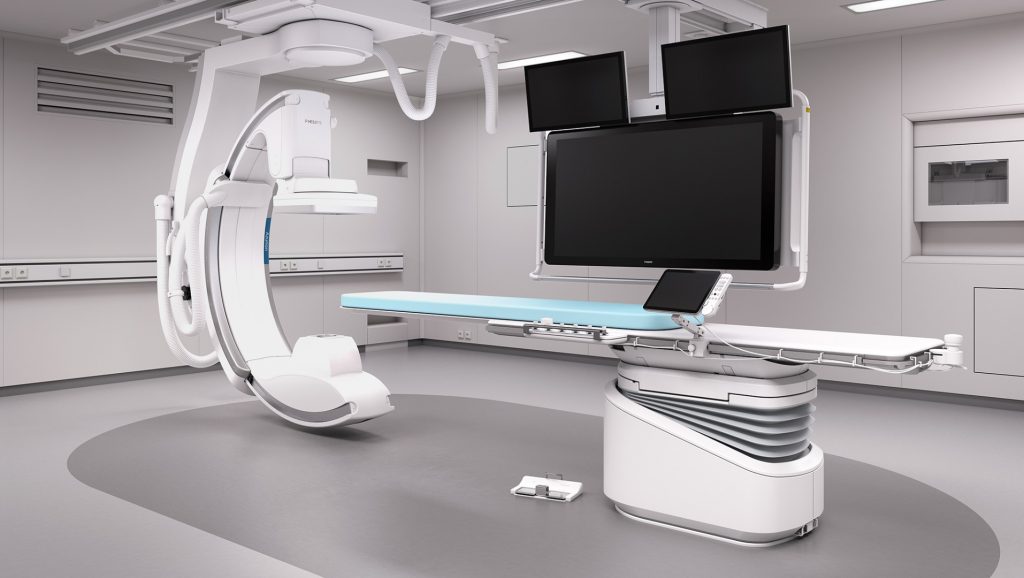 Philips Azurion 7, angiographic system
The angiographic complex helps to monitor the patient’s vessels in real time and monitor how the surgeon’s instrument moves through them. This is what such a system consists of:
- An irreplaceable part of the angiographic complex -C-Arm. The device was given this name because of its shape: it really resembles the letter “C”. It is the C-arc that includes an X-ray tube and a detector - a device that captures X-rays that “transparent” the human body. On the most advanced systems there may be several detectors. Modern C-arms can move in all planes, rotate 360 degrees and move on special rails on the ceiling or a tripod on the floor of the operating room. This allows timely scanning of the area of interest to the surgeon.
- A movable operating table on which the patient lies.
- High resolution monitor so the doctor can see the smallest details of the procedure.
- Intelligent software.It is this that turns the angiographic system into such a reliable and multifunctional complex. For example, special programs make hybrid imaging possible: if an X-ray image is not enough for a surgeon to accurately work, he can superimpose it on CT, ultrasound and MRI images obtained previously. Why is this necessary? The fact is that three studies - ultrasound, MRI and X-ray - allow you to see different types of human tissue. And by combining the images, the doctor gets the most complete picture.
One example of cardiovascular diseasessystems that can be treated with the help of an angiographic complex are aortic aneurysm, a common and life-threatening pathology. The aorta is the largest human vessel, and an aneurysm is an enlarged section of it with a weak, thin wall. The danger of the disease is that it can progress and lead to complications, the most dangerous of which is aortic rupture. Without urgent surgery, a rupture can cause death. To prevent the patient's condition from worsening, surgeons strengthen the vessel walls with special devices - polyester and metal tubes called stent grafts. The stent graft is inserted through a puncture in the femoral artery, and the surgeon “advances” it to the aneurysm under X-ray control. The device is then opened to the required size and finally implanted into the aortic wall. To ensure that the procedure is error-free and as safe as possible for the patient, surgeons can use technology for precise navigation within the vessels. An example of such technology is Philips VesselNavigator. This program automatically segments and marks areas of the aorta, which greatly simplifies the surgeon's work.
There are also innovations that will allowcreate an individual dynamic map of the coronary vessels for each patient (Dynamic Coronary Roadmap technology). The map is called dynamic because during surgery it can adapt to the movements of the patient's heart, allowing the doctor to act more accurately and confidently. Most importantly, it helps reduce the use of contrast media and operate on patients for whom such procedures were previously contraindicated due to chronic kidney disease.
How do technologies help prepare for minimally invasive heart and vascular surgery?
Planning for minimally invasive surgery - at leastan important stage than its implementation. The angiographic complex must be equipped with software that allows the surgeon to know in advance all the details of the upcoming intervention.
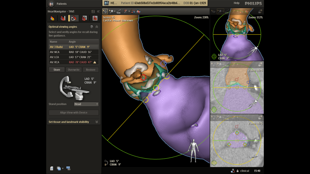 Philips HeartNavigator
The angiographic image is initially two-dimensional.But technology allows doctors to obtain a 3D picture. For example, the Philips HeartNavigator solution reconstructs 3D images from previously acquired 2D CT data sets. During surgery, the X-ray image is superimposed on the 3D data, which provides the doctor with additional information to improve the accuracy of the procedure.
Alexey Viktorovich Azarov, candidate of medical sciencesSciences, Head of the Department of Endovascular Treatment of Cardiovascular Diseases and Arrhythmias, Leading Researcher at the Moscow Regional Research Clinical Institute named after. M. F. Vladimirsky"
How is a laser used in minimally invasive surgeries?
A laser is a source of a very narrow and powerful beamSveta. The word “laser” is derived from the English abbreviation LASER - light amplification by stimulated emission of radiation, which means “light amplification by stimulated emission”. Laser radiation is highly directional and has high energy density. It is enough to act even on solid substances. The scope of laser application is very wide - from metal processing to information storage (an example of the latter is a DVD player). In medicine, a special type of laser is used - excimer. It is with its help that, for example, laser vision correction is carried out.
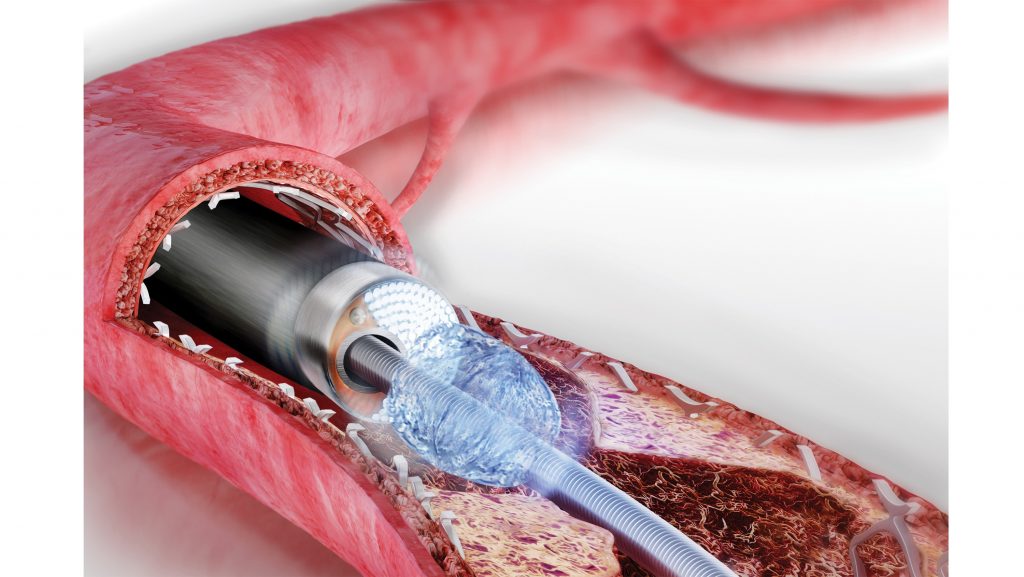 Laser application
The excimer laser is used in minimally invasivevascular surgery to combat atherosclerotic plaques and other pathologies that impede normal blood flow. The laser literally vaporizes the excess tissue in the vessel. To deliver the laser to the problem area, a special catheter is used that is connected to equipment that fires the beam. The first laser pulse reaches the tissue in 135 billionths of a second. The beam acts directly on the lesion inside the vessel and vaporizes five nanometers of tissue with each pulse. A few passes with a laser catheter are enough to remove the bulk of the atherosclerotic plaque.
Laser technology is more successful than itsalternatives. A number of clinical studies have shown that complications arising during operations of this kind are less than 3%, and the technical success of the procedure is 95–97%. This is a very high figure.
Modern technologies for minimally invasiveHeart surgery is a real revolution in healthcare. All the most important technological changes in this area have occurred in just the last couple of decades. New developments that make minimally invasive surgeries more accurate and safe appear every year. Today, some experts believe that approximately half of all heart surgeries can be performed without any incisions - and therefore with minimal risks for patients. This is a great example of how high technology improves the quality of life of millions of people.
Read more:
Einstein is wrong again and his main theory was rewritten: how it changes the world
Published test video of the world's first propeller with 11 blades
It turned out how old the water we drink today