How can you look into the brain?
The slide shows different brain imaging techniques. The first picture where
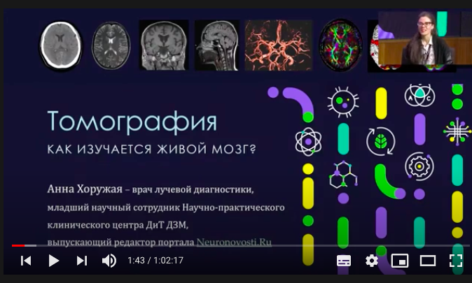
Then the interesting begins - we havethe ability to use blood as a contrast, so you can get angiography. This is a study of the vessels of the brain, which does not imply the introduction of any contrast agent from the outside, the contrast is human blood. So we can build a beautiful image of the vessels of the brain, and here the circle of Willis is visualized - the main circle of the collateral, that is, those vessels that communicate with each other and supply blood to all areas of the brain.
The following three color images renderstructural and functional tomography. And the image in the colors of the rainbow is magnetic resonance tractography, or diffusion tractography. It allows us to see how the tracts, the nerve pathways that come from each nerve cell, come together and go, for example, from the cerebral cortex down to the spinal cord and on to the muscles.
Penultimate images with bright orangestained is functional magnetic resonance imaging. This is one of the most interesting MRI techniques, which has a limited use in clinical practice, but it is widely used in scientific research. This method allows you to see the functional activity of different areas of the brain at the moment when a person is doing something or is at rest.
The latest image ispositron emission tomography, the most expensive method in radiation diagnostics, is actively used in some clinical situations. Here there is a radiopharmaceutical that is injected into the bloodstream of a person, then you can register the areas that will accumulate it the most.
CT scan
Scientists have a huge number of tools,which allow you to look into the brain, view the whole body. This is very useful in terms of clinical medicine and diagnosis by clinicians.
But what happened before that?How did clinicians arrive at a diagnosis by tapping, listening, and communicating with the patient? In 1896, there was a revolution in medicine - the X-ray was invented, it became enormously widespread. And then it began to be widely used in clinical practice.
Unfortunately, X-rays are activeaccumulates in bones, including the skull. Through this bright image, it is difficult to see the internal structures and what is behind the cranial box, it does not allow you to see the soft tissues of the brain. The first to find a solution to the problem was Walter Dandy. In the 1920s, he invented a method called ventriculography, around the same time pneumoencephalography appeared.

What it is?We cannot look through the bones of the skull into the brain, but we know that there are cavities inside the organ that are filled with cerebrospinal fluid, which is different in composition from blood, but, nevertheless, does not interact with X-rays. We can pump out this fluid, replace it with air or another fluid - and it tells us what is in the brain tissues.

A procedure where you need to pump out several dozenmilliliters of fluid from the system is very complex, closed, and the slightest fluctuations can cause fatal consequences. But researchers and doctors managed to do it. This method was the main method of brain imaging until the 1970s. Then Godfrey Hounsfield created a method that has now come to the fore in terms of diagnostic significance - this is computed tomography.
Pictured is a photo taken on October 1st.1971 - a snapshot of the brain of a living person. On it we can see a cyst filled with fluid. This shot was grainy and low quality, but even that was a colossal breakthrough. The first CT scan was taken around 1969. This is a picture of the brain of a dead young bull, Godfrey Hounsfield was setting up the technique on it.
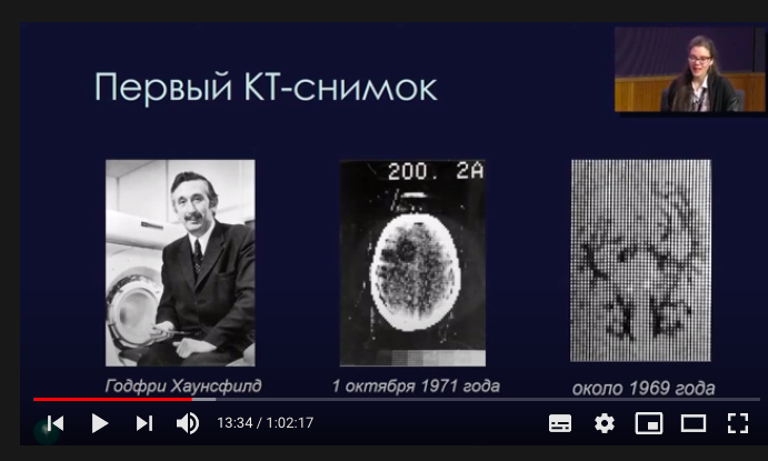
Interestingly, without the Beatles, developmentcomputed tomography would not be as active. In the 1960s, EMI, where Godfrey Hounsfield worked, was also a recording company. Thanks to a contract with a group gaining tremendous popularity, the funds appeared, on which Hounsfield improved computers, and they made it possible to process a large amount of information received from computed tomographs.
This is what the first CT scanner looked like at Atkinson Morley Hospital in London. And this is the same woman who was the first to undergo this procedure.

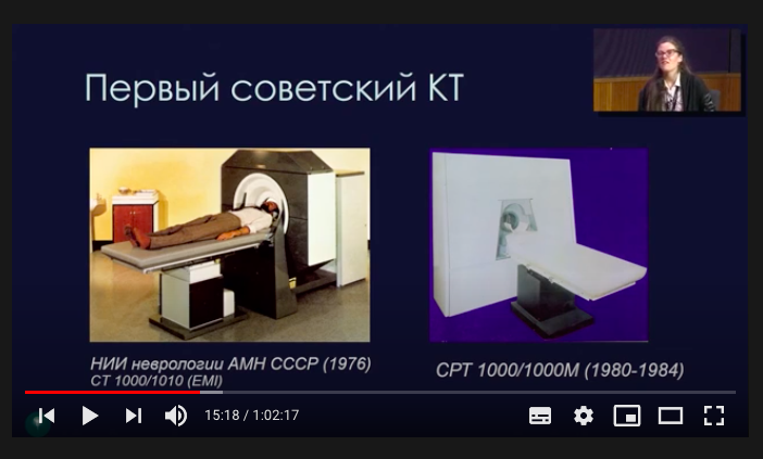
In our country, computed tomography begandevelop almost immediately after their appearance in the UK. The first CT scanner appeared at the Scientific Center for Neurology - this is my second alma mater, the place where I did my residency. I talked with the first X-ray laboratory assistant in our country, she worked on the first computer tomograph in the USSR.
She still works there and toldamazing stories: in the past, CT scans took so long that the patient had to lie still for hours in order to receive normal images of the brain. For example, one day she was distracted, and when she returned, she noticed that no one was in the scanning room. It turned out that the patient had already been lying there for two hours and he wanted to go to the toilet. It was returned and scanned for another hour or so. So research that lasts a few seconds is a big boon.
Positron emission tomography
Immediately after computed tomography appeared andpositron emission tomography. Its ancestor was the psychiatrist and neuropsychiatrist Louis Sokoloff. He figured out how to create a radiopharmaceutical and use it to visualize brain activity. Sokoloff worked during the war years in the United States and was very interested in understanding what happens in the brain of a soldier during a shell shock and how it then disappears.
But there were no such methods.Naturally, there was electroencephalography, which made it possible to measure the electrical activity of the cerebral cortex, but it could not move into deeper structures. The first positron emission tomography was done on August 16, 1976 on the brain.
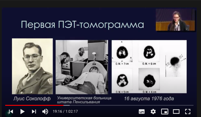
The black areas are the cerebral cortex.The first radiopharmaceutical was fluorodeoxyglucose. What is glucose - this is the main nutritional component for neurons, so the actively working nerve cells that make up the cortex actively absorbed it and signaled that they had a lot of mutated glucose. Therefore, we get an image of a bright black cerebral cortex.
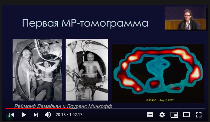
And this is the first magnetic resonance imaging.On the left, its creators are Raymond Damadian and Lawrence Minkoff. It was made on June 3, 1977. This method is fundamentally different from computed positron emission tomography. It does not contain ionizing radiation, it is absolutely safe.
CT scan
Already by the name of the method (other Greek.τομή - "section") it is clear that we are talking about the image of the section, the layer-by-layer measurement of the object's density by X-rays, followed by mathematical computer processing of the data. So you can get a three-dimensional picture without violating the integrity of the body. Information about each layer is collected into a single picture, it can be reconstructed into an image in any plane.
In this case, there is an x-ray sourceradiation - an x-ray tube, the researchers shine through the desired object. Depending on the density of the tissue, the X-ray radiation, as it were, hangs, remains in various tissues of the body. Bones have the highest density, they retain almost 100% of the radiation. The lowest is air. The data is collected in a detector, then they are converted into a digital image and the image that we see on the screen is built using algorithms.

There are several generations of devices, so farthere is a traditional computed tomography, which is now practically gone. There, the tube, together with the detector, circles clockwise, makes a full circle, and then the table advances a little. The tube makes another turn, and so on.
And the MSCT method is widely used.Here the table does not stop, it moves, and the tube with the detector rotates around the patient in a very dense spiral and illuminates the required area of the body in a fairly short time. This happens quickly, devices can make 256 and even 512 revolutions per second. But now, researchers are rather moving towards reducing radiation exposure and improving the quality of research.

The picture shows the result of a CT scan of the head. It shows that something is wrong - one of the hemispheres is clearly larger and slightly lower in signal intensity.
Computed tomography can alsolook at how blood is supplied to different areas of the brain, this method is called perfusion. And in the same patient, blue-blue shades can be seen. This means that the blood supply is impaired, we can conclude that a blood clot or embolus is stuck somewhere. Now some clinical actions can be taken with the patient.
In addition, there is a computed tomographyangiography, it is performed using a contrast agent. The contrast agent, by densely filling the vessels, can form a very bright picture, which we can evaluate by building three-dimensional images.
</ p>Magnetic resonance imaging
This method greatly expands the possibilitiesclinician and radiologist. This is the gold standard for brain imaging. It allows you to get images of internal organs in vivo, which are based on nuclear magnetic resonance. This is a phenomenon from the quantum world, so I will simplify some things so as not to dive into all the physical subtleties.
A permanent magnetic field is formed in the complex.The patient is placed there, where he stays for some time. A permanent magnetic field is formed there, it is 10 thousand times larger than the Earth's magnetic field, but this is not at all scary. There is no radiation in magnetic resonance imaging, this is one of the safest methods.
</ p>How does he work?Our body is mostly made up of water - two hydrogen atoms and one oxygen atom. Accordingly, hydrogen is the most common element in our body. Hydrogen and several other elements have certain physical properties - to simplify, they can rotate around their axis, that is, precess. These axes of rotation can look randomly in completely different directions.
Just placing a person in a strong magneticfield is not enough to receive any signal. We must influence the protons. This influence is handled by radio frequency beams, which are supplied by radio frequency coils.
Coils are additional add-ons inmagnetic resonance imaging. When a patient has an MRI of the head, an additional helmet is put on. These are coils, most often they are both receiving and transmitting. They can both emit a radio frequency pulse and catch a signal, that is, be a detector in order to catch the signal back.
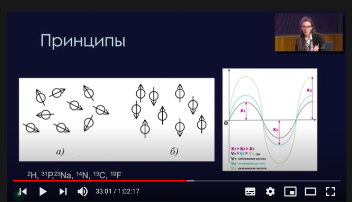
We affect protons with radio frequencyradiation at a frequency that is close to the rotation frequency of the proton and, thus, we deflect the arrow. We get a coiled spring, we give it energy, we can deflect it 90 or 180 degrees, depending on what we need. And when the RF pulse stops, the direction of rotation returns to the current position. Just like the spring that we compressed, it expands again to its original state, and energy is released, we call it relaxation, and this energy is recorded by the detectors that are located in the coils.
That is, the basic principles of MRI are to exciteprotons, atoms that we influence, then fix the relaxation, get the energy back, convert the figure into an image. This is also done by complex mathematical methods, such as the Fourier transform.
There are several generations of tomographs:for example, low-floor open. They are of the previous generation, the magnets are located above and below. Open machines are used in clinics because they are the only ones that can scan claustrophobic patients. There are high-field closed devices, where the maximum magnetic field strength is.
There are different modes of information collection in MRI - you canexclude elements or add information - for example, extrapolate an image a little. The first image is T2. Here you can see that the gray and white matter is rotated 180 degrees. This mode is needed because some pathologies are easier to see on a dark background. The second image is T1. On it you can see the anatomical structure of the brain, that is, the gray matter is really gray, the white is a little lighter.

There is another version of the image.This is a T2-weighted image with free fluid suppression. This is the same as the first one, but we removed the entire signal from the free fluid and got the opportunity to see the foci of the pathologically altered brain substance.
MRI can also be used to view blood vessels.Below is the angiography - the second image. We can look at the blood-brain barrier - this is the barrier between the blood and the substance of the brain, where it can pass and leak. Here, the area of the brightly glowing piece of the brain is edema, it tells us that this is where the ischemic stroke, the area of acute lack of oxygen, is located.
Functional MRI
This is the main method that is used in science.But it is also important for the clinical practice of neurosurgeons - if you need to remove a certain part of the brain, then you need to see if this will affect the function? To do this, a functional MRI is performed - preoperative mapping of the brain to see: how is the area located, for example, near the tumor that needs to be removed, and the area of the functionally active area of the cerebral cortex, for example, the speech center, and whether we will remove, for example, area of the speech center together with the tumor.
Using fMRI, you can capture, receiveauditory activation, that is, to see which areas of the brain are activated in response to sound exposure. You can get motor activation, for example, you can ask the patient to move a finger and fix the activity in the cortex that the movement caused.

You can also look at an inactive brain, becausethat he, too, spends a lot of energy maintaining his balance. In the picture, one of the most interesting networks is the network of the passive mode of the brain. It is believed that this network partially reflects the presence of human consciousness. Scientific research in the field of consciousness is one of the most ambitious things in the field of neuroscience.
Traktografiya allows you to fix the movementprotons along axons, nerve pathways. So we can get beautiful images, here each color is encoded with a direction. From these colors you can get very important information. This is necessary in clinical practice, for example, during a neurosurgical operation, so as not to touch a strategically important piece of this highway. This is how the program in which you can build tractographs looks like.
Positron emission tomograph
This is a radionuclide method for studying internalhuman organs, where antimatter is formed and annihilation occurs. These are difficult words, but they can be found in the novels of Dan Brown. From them, we remember that even a small amount of antimatter mixed with matter is enough to wipe a city from the face of the Earth. But this method should not be feared, it can bring a relatively small amount of radiation, which is within the normal range.
What is the principle of positron emission tomography?The fact that the half-life of fluorine-18 is 110 minutes, so you need to have time, firstly, to synthesize a radiopharmaceutical, and secondly, bring it to the clinic, where it will be administered to the patient, wait until all this glucose has spread throughout the patient's body, then take pictures. However, fluorine decays via beta-plus decay and releases a positron. It meets the first electron it comes across, interacts, annihilation occurs, and two gamma quanta are detected by detectors. This way, researchers get the brightest image possible where most of the radiopharmaceutical accumulates.

This is what hybrid research looks likecombine PET-CT, PET-MRI, this is now one of the new methods. At the same time, there is also a combination of functional activity and structural activity to obtain clinical information. Not so long ago, a whole body PET scanner appeared - this also provides a lot of interesting and clinically significant information. From the point of view of innovation and technology, science can still develop forward, and in many areas - CT, MRI, PET - and make scientific, scientific and technical improvements there and contribute to the creation of new technological and high-tech medicine.
Read more
Look at the "silent" drone with a new generation of ion propulsion
Ancient trilobite males strapped females on during mating
Russia and the United States have Doomsday planes: how and where they will fly in the event of the end of the world