Research team led by University of Freiburg professor Alexander Rohrbach
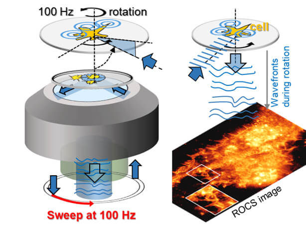
The laser rotates around the object under investigation at different angles 100 times per second. Every ten ms, an ultra-high-definition image is formed based on scattered light.
 Source: Rohrbach, University of Freiburg
“We use several physical phenomena,known from everyday life, says Rohrbach. “First of all, the fact that small objects, such as molecules, viruses or cellular structures, scatter blue light the most.”
This specificity of tiny objects, as notedscientists, it is easy to show on the example of the sky. Air molecules scatter the blue part of the solar spectrum the most, which is why the daytime sky appears blue to us. In the context of microscopy, small objects, according to the authors of the development, scatter and direct into the camera about ten times more blue light particles than red light particles.
The second feature, also borrowed fromreal world, the angle of inclination at which the beam is directed towards the object under study has become very low. The researchers say that images of particles become clearer when the laser beam is tilted to the plane of the object, just as fingerprints are more visible on a glass when viewed at an angle to the light.
In addition, scientists illuminate the object with an oblique laser beam sequentially from all sides to avoid possible distortions and artifacts.
ROCS uses blue, collimated laser light rotatingunder oblique angles to form images within 10 ms. Hence, back scattered laser light forms a super-resolved image on a camera within 10 ms by simply adding up coherent images (left movie part). Right: image formation with 700x slowmo pic.twitter.com/JBcfBLfSec
— Alexander Rohrbach (@AlexRohrbach09) January 3, 2022
On the left - individual images, on the right - the overall image.
Researchers demonstrate workmicroscope on various cell systems. For example, scientists have been able to film how stimulated mast cells open small pores in just a few milliseconds to shoot spherical pellets with inexplicably high force and speed. The granules contain the messenger histamine, which can subsequently lead to allergic reactions.
In other experiments, scientists were able to observemany thousands of images of how filopodia — the long, filamentous “fingers” of macrophages — scan their environment for prey in a complex quivering motion, and how their cytoskeleton can change at previously unknown rates.
Amazing how fast virus-like (100nm, n=1.4) particles are, how they try to find the best binding point at the cells (100 Hz ROCS microscopy, 5x slomo) pic.twitter.com/04yGMyWSkQ
— Alexander Rohrbach (@AlexRohrbach09) January 2, 2022
Virus-like particles try to enter the cell
“Our main goal was not to create beautiful images or films with unexpectedly high cell dynamics – we wanted to gain new biological knowledge,” says Rohrbach.
Read more:
MIT builds stationary heat engine that outperforms turbines
After ten years of work, scientists questioned the standard model of physics
See what sunrise looks like on Mars