How did the technology of radiation diagnosis
The starting point in the history of radiological diagnostics is
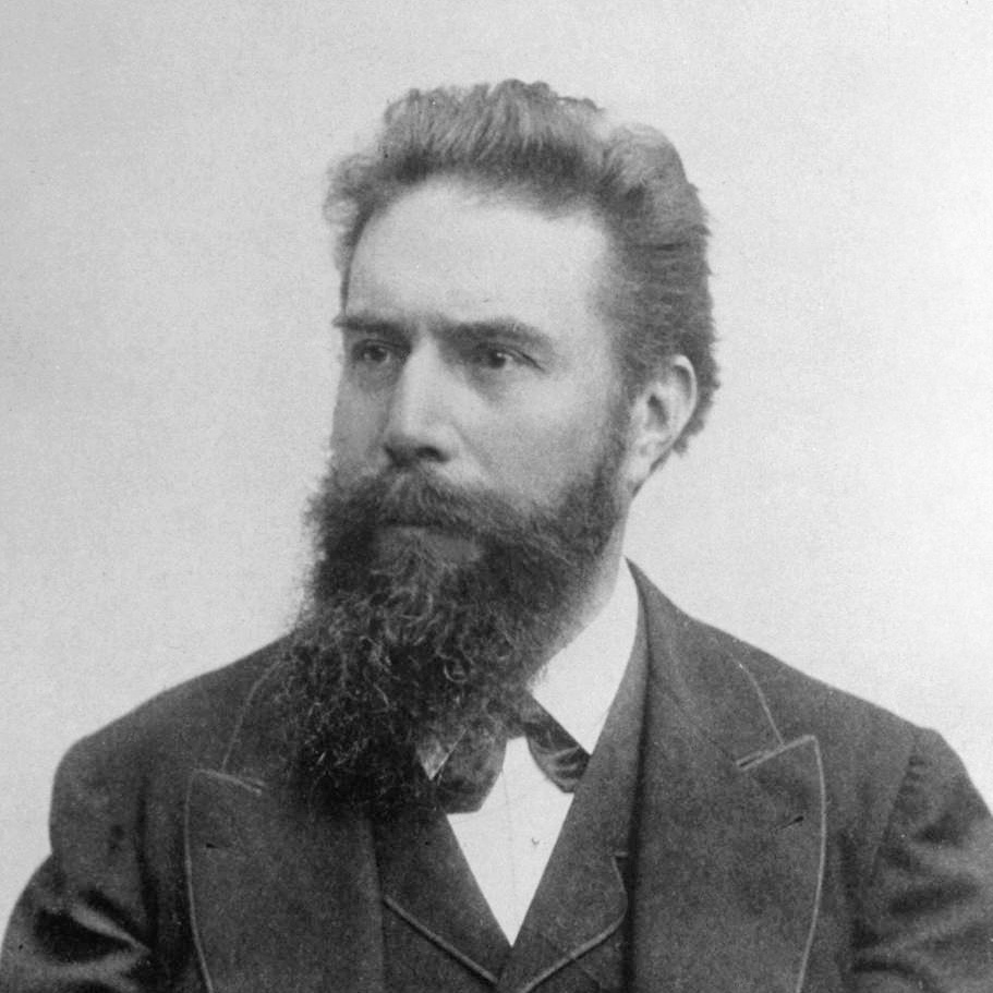
Wilhelm Konrad X-ray- German physicist from the University of Würzburg.During his career, he also worked as a professor of physics in Hohenheim, Strasbourg, Giessen and Munich. The first Nobel Prize laureate in the history of physics (1901).
X-ray was investigating piezoelectric andthe pyroelectric properties of crystals, established the relationship of electrical and optical phenomena in crystals, conducted research on magnetism, which served as one of the foundations of the electronic theory of Hendrik Lorentz.
But the main discovery of X-rays was the X-rays,which he discovered when he was already 50 years old. Representatives of industrial firms repeatedly approached a scientist with proposals for a bargain purchase of rights to use the invention. But Roentgen refused to patent the discovery, because he did not consider his research a source of income.
For evil irony - X-rays died from cancer, in the modern diagnosis of which the invention of the scientist is actively used today.
Several German companies have been working onthe creation of X-ray tubes, but they were too expensive, and many clinics could not afford this equipment. In 1918, Philips developed its first medical X-ray tube, which made a breakthrough in the fight against tuberculosis and allowed doctors to take control of the spread of the disease. The principle of X-ray operation has not changed over the years: an X-ray tube generates radiation that passes through the human body and hits the detector. Different tissues transmit or block X-rays differently, resulting in an image being formed.
 Wilhelm Konrad X-ray
In 1932, a reduced anda lightweight version of the X-ray machine that was used, including the doctors of the Dutch army. Already by 1939, it was released 100 thousand vehicles. X-ray tubes began to be delivered to medical institutions around the world due to an affordable price, and the spread of knowledge about X-rays influenced the creation of the first CT scanners.
How bats helped doctors
The history of ultrasound began in the 18th century, whenItalian physicist and naturalist Lazzaro Spallanzani drew attention to the ability of bats to navigate in complete darkness. Empirically, the scientist found that even the lack of vision does not interfere with this. But wax plugs in the ears caused nocturnal animals to lose orientation in space. Spallanzani suggested that bats make a certain sound, inaudible to humans, which is reflected from surfaces and helps the animals to easily avoid obstacles. At that time, ultrasonic signals could not be detected, so the scientist’s assumptions remained hypotheses.
 Bats orient themselves in total darkness due to ultrasound reflected from surfaces.
In 1880, physicists Pierre and Jacques Curie found thatSome crystals (for example, quartz) are capable of recognizing an electric field when subjected to mechanical action. Thanks to this discovery, the first ultrasonic detectors were created - the main elements of devices that, for the first time, made it possible to technically receive an ultrasound signal.
Equipment, vaguely reminiscent of modernmedical devices, was created only in the 1950s. English surgeon John Julian Wilde first measured the thickness of the intestinal wall with ultrasound, developed special sensors for diagnostics, and also found out that malignant tissue reflects ultrasound waves better than healthy ones. The prototype of the modern ultrasound device, where the sensor is in the doctor's hand, appeared in the United States in 1963 - from that moment ultrasound was widely included in medical practice. Today it is the most accessible and safest diagnostic method and is used everywhere to study cardiovascular, oncological diseases, the work of the gastrointestinal tract and others.
Safety and reliability - MRI discovery
In 1946, Felix Bloch of StanfordUniversity and Edward Purcell from Harvard independently discovered the phenomenon of nuclear magnetic resonance, for which both were awarded the Nobel Prize in Physics. Few people know that the first scientist who, back in 1960, proposed the use of magnetic resonance imaging (MRI) for diagnosing diseases was the Soviet scientist Vladislav Aleksandrovich Ivanov.
Despite this fact, the founding date of MRIIt is generally accepted that 1973 was the year when Professor of Chemistry and Radiology Paul Lauterbur published the article “Image Creation by Induced Local Interaction” in the journal Nature; examples based on magnetic resonance." This work formed the basis of MRI diagnostics.
 MRI provides cut-through images of organs using magnetic resonance
Another scientist Peter Mansfield improvedmathematical imaging algorithms. Later, there was a way to get short-cut images of organs using magnetic resonance. Simply put, visualize the state of the internal organs and tissues of a person by placing it in a strong magnetic field. In 2003, Lauterbur and Mansfield were awarded the Nobel Prize in Physiology and Medicine for this discovery.
2-in-1 Diagnostics: Discovery of the PET / CT Method
Positron emission tomography (PET) was performeda long development path from use in a scientific laboratory to implementation in clinical practice. Thanks to new discoveries in this field in the 50s, doctors were able to see the distribution of a radiopharmaceutical - a biologically active compound labeled with a radioactive atom - in the human body.
The first prototype of a PET scanner appeared in 1952at Massachusetts General Hospital, but with its help doctors received only one two-dimensional image, and not a sequence of them. This was due to the fact that the scanner had only two detectors, located to the left and right of the patient's head, and the resolution was low. However, the sensitivity of the device was still able to detect the tumor.
 Image constructed by the method of projections of maximum intensity
In the further improvement of PET went on twodirections: the number and location of sensors increased, in parallel with this developed methods of mathematical data processing. In the late 1970s, PET scanners began to be widely used in clinical practice, and in the early 90s, an oncosurgeon, Rudi Egeli of the University of Geneva, suggested placing CT equipment in the gaps between the PET scanner sensors in order to receive and metabolism in the patient's body. This is how the combined PET / CT scanners appeared, which are now used in modern clinics around the world.
Innovation here and now
It took only half a century to visualize inmedicine has jumped in its development. Full scan of a person in one click, the possibility of transferring images over a distance and remote consultations with other experts - could Wilhelm Roentgen or Lazzaro Spallanzani dream of this? Today, developments in the field of radiation diagnostics are aimed at improving the quality of visualization, since a clear image allows doctors to conduct an accurate examination, make the correct diagnosis the first time and quickly determine further treatment tactics. Moreover, modern technologies help to make new research and breakthrough discoveries not only in medicine, but also in other fields, for example, in archeology and neurolinguistics.
CT scanner guarding health and helping archaeologists
The heart is the only organ in the body thatis in continuous motion. When we shoot an object in motion with a camera, it turns out blurry and unclear. But modern computed tomography preserves the clarity of the image. Today, CT scanners make it possible to examine not only the heart in a few seconds, but also obtain high-precision images of blood vessels, the bone skeleton and other organs.
Philips specialists went even further anda few years ago created the spectral computer tomograph IQon. This device has become the first system in the world that works on the basis of a unique dual-layer detector. It simultaneously distinguishes photons of X-ray radiation of high and low energy levels, which makes it possible to obtain not only anatomical information, but also data on the composition of tissues. This helps physicians make more informed decisions on the further tactics of diagnosis and treatment of the patient. After conducting a study on spectral CT, radiologists can analyze objects that are indistinguishable by conventional CT scanning.
Sidebar
CT helps not only to conduct a surveyman, but also opens the veil of secrets of ancient culture and history. A few years ago, thanks to the tomography capabilities, the specialists from Philips and the Naturalis Museum managed to look into the past 66 million years ago and study the caudal vertebrae of the Rex Rex. And before that, with the help of Philips CT, studies were carried out on the remains of the inhabitants of the city of Pompeii, which was destroyed during a catastrophic volcanic eruption in 79 AD
Tumor under the gun: what digital PET / CT is capable of
PET / CT combines two types at onceresearch: computed tomography evaluates the structure of the modified tissues and helps to determine their location to the millimeter, and positron emission tomography creates high-precision three-dimensional images that allow you to see the processes occurring in tissues and organs.
Sidebar
Today, PET allows doctors to identifytumors are from three millimeters in size, and CT is capable of determining its location with millimeter accuracy. This method has become a real revolution in medicine: with the help of PET/CT, doctors can diagnose the brain, identify age-related diseases - Alzheimer's and Parkinson's diseases, and also recognize coronary heart diseases. In addition, PET/CT helps to detect the presence of cancer cells in the early stages and, if necessary, quickly begin treatment.
With this method, the surgeon will know exactlywhere is the tumor, what is the dynamics of its development, which will provide an opportunity to completely remove it without affecting healthy organs. Also, experts can understand how best to conduct radiation therapy to kill cancer cells with minimal damage to healthy tissue. Recently, Philips developers have introduced the Vereos full digital PET / CT scanner to the Russian market, which uses a digital detector instead of traditional photomultipliers. The device catches even very low doses of radiation while maintaining high image quality, which is safer for the patient and the doctor. The equipment allows you to control radiation exposure without losing visualization quality and maintain the clarity of images even in the presence of implants.
What's new in the field of MRI and where is neurolinguistics
MRI diagnostics today is considered quitea standard procedure that can be performed in many medical centers. But few people know that MRI is a valuable tool not only for doctors, but also for specialists in the field of neurolinguistics.
What happens in our head when we hearspeech or something we say? How to help people with speech pathologies? MRI helps professionals to “see” the language. So, scientists from the HSE laboratory of neurolinguistics use functional MRI to study adults with various brain lesions affecting speech function. Thanks to this diagnosis, you can see the damaged area, and how the brain builds new ones instead of broken links. According to MRI data, one can understand where the speech function has gone. Scientists have also developed a special speech localizer: people perform a speech task in MRI, with the help of which the activity of brain regions is determined. Based on the data obtained, optimal therapy is selected.
Another example of MRI is non-invasive.assessment of the concentration of iron in the human body. This study was conducted by specialists from the Research Institute. Rogachev. Children suffering from blood diseases receive it from donors, which over time leads to the accumulation in the tissues of iron-containing compounds - the breakdown products of blood hemoglobin. This leads to serious violations of the functions of organs, for example, in the heart - to a sudden stop and cardiomyopathy, in the liver - to cirrhosis, in the pancreas - to diabetes. Usually, the control of the concentration of iron is checked using a liver biopsy, but this method is invasive and can lead to serious consequences. Modern methods of MRI allow, without surgical interventions, to assess the concentration of iron in the tissues; however, additional research is still required to introduce the method into clinical practice.
Cardiomyopathy- a heterogeneous group of myocardial diseases,associated with mechanical or electrical dysfunction, which usually manifests as inappropriate hypertrophy or dilatation. Cardiomyopathies can either affect only the heart in isolation or be part of a generalized systemic disease, often leading to cardiovascular death or disability caused by progressive heart failure.
Medical diagnostic equipment passeda great way and improved every year, allowing you to get better images. In September 2018, Philips revolutionized the industry and introduced the first MRI machine in Europe with a revolutionary cooling system to achieve the effect of superconductivity. Unlike the classic magnet, which requires more than 1,500 liters of liquid helium to cool, only 7 liters of this liquefied gas is involved in the new device. Liquid helium is placed in the system at the manufacturing stage, after which the magnet is completely sealed, which eliminates the possibility of gas evaporation and eliminates the need for regular refueling. The gelless scanner is simpler to install and allows you to significantly reduce the cost of operation of the clinic.
Step into the future: what discoveries are worth waiting for in the XXI century
With the development of digital integrated technologyTeleradiology capabilities will gradually expand. The essence of this sphere consists in the exchange of diagnostic images and other patient data inside and outside the clinic for remote conclusion or for obtaining a second expert opinion. With the help of teleradiological systems, it is possible to improve the quality and availability of medical care to the population anywhere in the world.
To the aid of doctors in the analysis and description of medicalimage gradually comes artificial intelligence. AI provides a comprehensive analysis of all the information available in the snapshot, thereby reducing the risk of accidental omission of pathology that was not in the field of view of a specialist. Also, the AI will solve the problem of the quality of medical images, because it can automatically check if there are any defects in the image. Due to this, the number of repeated studies is reduced, and clinics can more effectively allocate the budget due to this. Philips already has a similar prototype solution.
Sidebar
Great impetus to the development of radiation diagnosiswill provide augmented reality technology. One of such breakthrough solutions is the development of Voka, which allows the traumatologist to see damaged bones inside the patient at the examination and planning stage of the operation. Based on the data obtained as a result of CT or MRI, 3D-models of injured organs and tissues are created. The resulting models, as well as models of implants and spokes, are loaded onto the Microsoft HoloLens mixed reality headset for further work of the surgeon. This ensures high accuracy of operations and rapid rehabilitation of patients even after serious injuries. A similar concept was developed by Philips specialists together with Microsoft for HoloLens 2. The solution allows real-time transfer of 2D images to a three-dimensional holographic environment of augmented reality, which can be easily and intuitively controlled by a doctor. This concept is specifically designed for minimally invasive operations, where accurate and detailed visualization is the key to a successful procedure.
Big data will play a huge role. On the one hand, they contain huge arrays of information about patients, and therefore allow you to store more knowledge about existing pathologies. This will contribute to an earlier and more accurate diagnosis of not only diseases, but also various predispositions. On the other hand, big data will help to create libraries of structural data, which will allow scientists to get an answer even before conducting fundamental and expensive research: for example, should the theory be tested and developed in the development of a new drug.
These 4 spheres are key aspects of the radialdiagnostics that will turn the world of radiation diagnostics and open up new opportunities for doctors in the fight against even the most serious diseases of the twenty-first century.