A monumental leap forward in stem cell research – experiment led by scientists from
All placental mammals (humans, mice, mooseetc.) start life approximately the same. Soon after fertilization, the first cell divides until three main areas of tissue are formed: one serves to create the animal itself, and the other two – for organs that promote its growth inside the mother.
If the former can independently create a modelembryo (or embryoid), then the presence of two second groups of placental cells nearby provides the necessary chemical "negotiations" that contribute to many of the smallest changes in the developing animal.
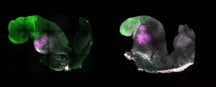
By mixing stem cells representing these threemajor tissue groups, and by refining previous methods for developing them in vitro into an embryoid, the team found that their model (pictured right) could develop “on its own”, developing a nervous system equivalent to a natural mouse embryo (pictured left) by 8.5 day after conception.
The synthetic embryoid also contained the mainheart tissue that was beating, and the rudiments of the intestines, as well as the beginning of structures that, in a real embryo, could build parts of the skeleton, muscles and other tissues under the skin.
By itself, the model will not continue to evolve into something like a mouse. Science is still far from being able to create a full-fledged organ from stem cells, not to mention a whole animal.
The presence of a collection of fabrics that reliably reflectdevelopment outside the body gives researchers the opportunity to not only observe but ethically test genetic changes that can help improve our understanding of how our bodies grow.