Researchers first used ultra-high-resolution MRI (7 Tesla tomograph) for comparison
Scientists studied the data of tomograms of people withchronic migraine, with episodic migraine without aura and a control group of "healthy" people of the same age. All participants were between 25 and 60 years old at the time of the study, and none suffered from overt cognitive impairment or a brain tumor.
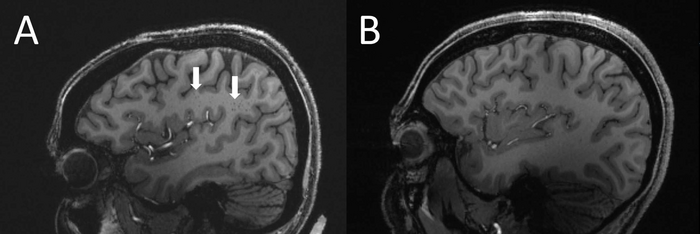 Enlarged perivascular spaces in a patient with migraine (left) compared to controls (right). Image: RSNA, Wilson Xu
Statistical analysis showed that the numberdilated perivascular spaces in the semioval center was significantly higher in patients with migraine compared with the control group. The researchers note that dilated perivascular spaces may be a sign of small vessel disease.
The perivascular spaces are part offluid excretion systems in the brain. Learning how they contribute to migraine will help us better understand the complexities of migraine.
Wilson Xu, New York University researcher and co-author
The researchers suggest that significantDifferences in the perivascular spaces between migraine patients and controls may indicate dysfunction of the lymph nodes in the brain. Although it is not yet known whether such changes are a cause or a consequence of migraines, future research will help establish this and develop an effective solution to prevent migraines, the scientists say.
Read more:
A magnetic storm is about to hit Earth
The true meaning of mummification is revealed: all this time, scientists were wrong
Named the main danger of the lunar mission "Artemis"