Researchers from Innopolis University, Kazan State Medical University and
In an experiment described in an article in the Journal ofDigital Imaging, four practicing radiologists with professional experience from three to 30 years participated. During the study, scientists recorded how the doctor’s gaze moves when studying an x-ray. The primary analysis shows how long it takes the specialist to evaluate the image and which areas attract the doctor's attention. Further analysis using AI revealed key patterns.
 Workplace of a radiologist equipped with an eye tracking system for conducting an experiment. Image: Innopolis University
Radiologists were asked to analyze 400images of the chest organs on a special device with an additionally installed system for eye tracking. An algorithm developed at Innopolis University based on a modified architecture of the U-Net neural network identified the contour of the lungs on chest x-rays and measured the area of the lung image that the doctor looked at.
On average, radiologists examined 55 to 65% oflung area on x-ray. The experiment showed that the more images doctors interpreted, the smaller part of the lungs they covered with a look, and the most experienced radiologist with thirty years of experience demonstrated the smallest area of coverage of the lungs with a look.
Ilya Pershin, Research Engineer, Laboratory of Artificial Intelligence in Medicine, Innopolis University
The study found that for every 100of the studied images, the area of the radiograph, to which the doctor pays attention, decreased by an average of 1.3–7.6%. The effect of fatigue on X-ray analysis was observed in all doctors and various pathologies in the images, but in different ways: in the presence of signs of dangerous changes, even “tired” radiologists studied the images more carefully.
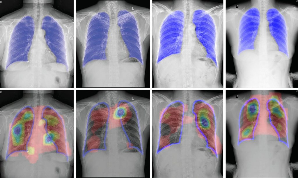 An example of assessing the areas on which the eyes of doctors were fixed. Image: Innopolis University
The authors of the study note that the missingpathology in the picture is one of the most common medical errors of radiologists. Understanding how fatigue and other factors affect the quality of image analysis will help optimize AI systems for image analysis and support radiologists with more information to make an accurate diagnosis.
Read more:
The mystery of the red stripes on the satellite of Jupiter is revealed
Migraines and headaches found a new explanation
Found "impossible" planet. She defies modern science