In order to study the organ on which surgery is to be performed, the surgeon evaluates the results of computer
To simplify the surgeon's work, specialistsThe NTI Competence Center in the field of Robotics and Mechatronics Component Technologies at Innopolis University has developed the HLOIA product. It can turn an organ into a 3D model using video and images based on MRI data.
Next, the information is saved in cloud storage, and then, using a new program, it uploads for mixed reality glasses, the necessary models and files for use during the operation.
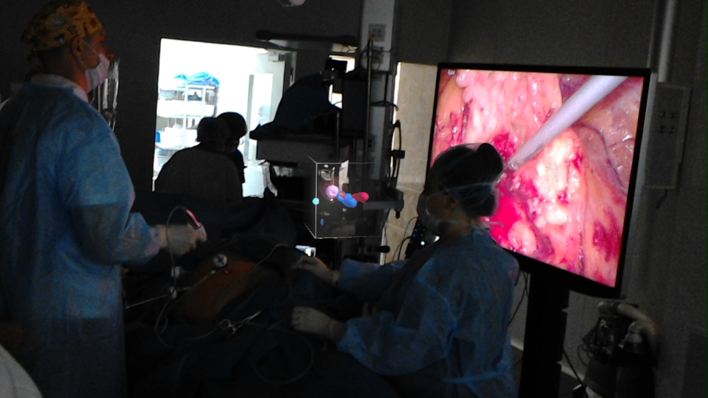
The glasses can be used in the operating room and withusing gestures, launch the application to re-observe the organ model, enlarging and rotating it 360 degrees - this helps to navigate during preparation and during laparoscopic surgery, when all manipulations are performed under the skin through small incisions.
Setting up the glasses takes a couple of minutes. The same algorithm is possible for any operation.
The first operations were performed by urologists on patients withlocalized kidney cancer with 1st generation Microsoft HoloLens mixed reality glasses, then switched to 2nd generation Microsoft HoloLens. In February 2020, the development was tested during laparoscopic pancreatic surgery.
Read more:
Unknown animals resembling sponges found in the ice of Antarctica
The Brunt shelf in Antarctica is collapsing at a speed of 5 meters per day
Abortion and science: what will happen to the children who will give birth