Researchers from the Max Planck Institute for Behavioral Neurobiology have developed a miniature
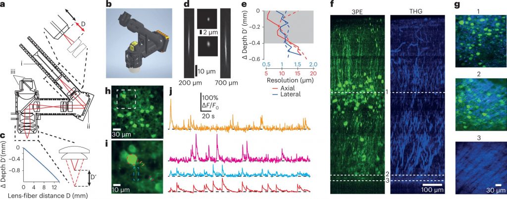 Scheme and model of the microscope and observational data. Image: Alexandr Klioutchnikov et al., Nature Methods
To understand how complex behavior is formed,it is necessary to conduct observations in natural conditions, the scientists explain. The new device works remotely, does not interfere with the free movement of animals and can analyze brain activity in the process of interacting with the environment.
The device is a three-photonforehead microscope. It weighs only two grams and yet records the activity of neurons with a resolution of one cell in all layers of the cerebral cortex. Since the focusing is controlled remotely, the behavior of the animal during the measurements does not change. Unlike analogues, the device can operate in illuminated conditions, and the modular design of the microscope provides the possibility of functional visualization with high resolution of neuron bodies up to their processes, dendrites.
To test the operation of the device, the researchersconducted measurements in the fourth and sixth deep layers of the cerebral cortex of mice. During the experiment, the experimental animals freely explored the space. The scientists found that nerve cells in different layers modulated differently, depending on how bright or dark the environment was.
This is a huge step to analyze brain activity deep in the cerebral cortex while the animal exhibits natural visually guided behavior.
Jason Kerr, Head of Brain Organization and Behavior at the Max Planck Institute for Behavioral Neuroscience
Read more:
Does science exist in extreme conditions? We answer in numbers
The egg was dropped from space: look what happened to it
Restored appearance of a medieval woman who suffered from syphilis
On the cover: an artistic illustration of a three-photon microscope. studying neurons. Image: Julia Kuhl, Max-Planck-Gesellschaft