In a new study, scientists from the University of Southern California created memories for the first time in
How are memories created?
While many aspects of how the brain works for the most partnot studied, scientists agree on how memories appear. They are thought to occur when certain clusters of neurons are reactivated. In general, experts agree that the brain creates memories by changing synapses (connections between neurons). However, this may not be the case.
In a new study, American scientistsfound that learning doesn't just change synapses, but causes them to grow in some areas of the brain and die in others. Biologists believe these changes will help explain how memories are formed and why some are stronger than others.
New research method
In the course of the study, scientists for the first time evaluated the strength andthe position of synapses in the brain of a living zebrafish. This model organism is often used to study the functions of the human brain. What's more, they compared synapses in the same brain over time, keeping fish alive and subjecting them to non-invasive interventions.
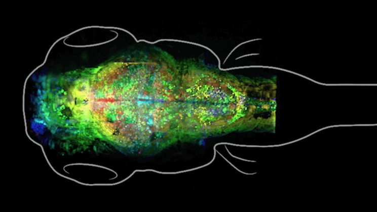 The brain of a zebrafish. Photo: dornsife.usc.edu
For the experiment, scientists have developed a new methodworking with data. Previously, researchers had been unable to determine the exact location of a synapse in a living brain without changing its structure and function. This means that it was simply impossible to correctly compare the state of the brain “before” and “after” the appearance of memories. Now the scientists used a new type of cell marking and a custom microscope made by Caltech engineers.
How was the study
During the study, scientists forced the fish(who was 12 days old at that time) “linked” the turning on of the light with the heating of the head by an IR laser, which caused them unpleasant sensations. Individuals that “realized” that turning on the light led to the appearance of a laser waved their tails in an attempt to swim away. This was a signal to scientists - the fish had successfully completed the training.
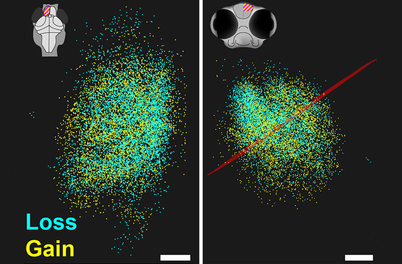 Using software developedResearchers at the University of Southern California, which creates a map of brain synapses from a 3D microscopic image, compared the size and location of synapses before and after training. The red line marks the border between brain regions. Image: Don Arnold
As a result, biologists managed to fixlarge-scale and unusual changes in the brain of fish. Instead of changing the strength of existing synapses, the scientists watched as connections between neurons were destroyed in one part of the brain and made in another.
main conclusion
The discovery changes scientists' understanding of how the brain works and how memories are formed. This will be useful to specialists in the field of neurology, psychology and psychiatry.
During the study, we focused onassociative memories. They are more stable than others and form in a different place in the brain, the amygdala, rather than the hippocampus. This will be useful in the treatment of post-traumatic stress disorder, which is thought to be related to the formation of associative memories.
Textresearch
As a result, the experiment clearly shows hownegative associative memories are formed and why they are so stable, affecting a person all his life. However, scientists believe that more research is needed. The goal is to understand how long associational negative memories and synaptic changes persist in zebrafish. The experts then want to see if these findings apply to animals with large brains.
Read more
"James Webb" took the clearest photo of a star in history
At a depth of 8,000 meters, scientists have found strange bacteria: they were not expected to be seen there
Scientists stumble upon 1,500-year-old archaeological anomaly